| |
A comparison in the progression of liver fibrosis in chronic hepatitis C between persistently normal and elevated transaminase
|
| |
| |
At the end of this report is study of interferon plus ribavirin in HCV+ patients with normal ALT presented at DDW 2002 by Ira Jacobson, MD.
Journal of Hepatology 38 (2003) 511-517
Chee-Kin Hui, Tigist Belaye, Kevin Montegrande, Teresa Lyn Wright *
Department of Medicine, GI Research (111B), Veterans Affairs Medical Center, 4150 Clement Street, San Francisco, CA 94121, USA
An estimated 20-30% of patients with chronic hepatitis C (HCV) infection have persistently normal alanine transaminase (PNALT). Studies have suggested that these patients have only mild hepatitis and that they may either not progress to cirrhosis at all or progress at a much slower rate than those with elevated serum alanine transaminase (ALT) levels. However, studies performed in those with PNALT have highlighted several characteristics of these so called
ÔasymptomaticÕ HCV carriers. For example, the presence of serum HCV RNA is almost invariably associated with some degree of liver damage despite PNALT
levels, challenging the very concept of ÔhealthyÕ HCV RNA carriers. In fact, Puoti, et al. found that 26% of their subject with PNALT had moderate to severe hepatitis whereas only 6% of those with raised ALT had moderate to
severe hepatitis. This study coupled with othersÕ, have led to controversies concerning the histological findings in patients with PNALT as liver cirrhosis occurred in 5-20% of these patients suggesting that the clinical course
of PNALT is not necessarily benign.
One clinical issue is whether fibrosis progresses in patients with PNALT at the same rate as does the fibrosis in patients with elevated ALT. Mathurin, et al. conducted a cross-sectional study to address this question but his study was based on a rate of fibrosis progression calculated from the presumed duration of infection rather than a direct histological comparison between patients with PNALT and those with raised ALT. A second study by Martinot-Peignoux, et al. compared PNALT patients with detectable HCV RNA to those with undetectable HCV RNA, and found more mild liver disease in the latter group, but did not include a compar-ison group of patients with elevated serum ALT. Since it is now known that fibrosis progression in HCV is non-linear, cross-sectional studies to determine fibrosis progression may not be accurate. We have therefore, conducted a study to compare directly the progression of fibrosis in patients with PNALT to those with elevated ALT, to compare the development of severe fibrosis in patients with PNALT and elevated ALT and to compare the long-term
outcome of patients with PNALT and elevated ALT.
Abstract. Detectable serum hepatitis C virus (HCV) RNA in HCV patients with persistently normal alanine transaminase (PNALT) has been found to be associated with significant liver damage. The primary outcome of this study was to compare the histological progression of fibrosis in patients with PNALT and elevated alanine transaminase (ALT). Forty patients with PNALT (Group 1) and 41 patients with elevated ALT (Group 2) were recruited into this study. Only patients with fibrosis of F0 to F2 were recruited into this study. The median time to second liver biopsies was 6.3 (range 2.0-11.1) years. Nine patients (22.5%) in Group 1 and 17 patients (41.5%) from Group 2 had progression of fibrosis. There was a trend towards a significantly higher cumulative probability of fibrosis progression in Group 2 (P =0.06). In patients with an initial F0 to F1 fibrosis, there was a significant difference in cumulative probability of fibrosis progression between Groups 1 and 2 (22.6% (7/31) vs.
43.3% (13/31), respectively, P =0.02). Anti-HCV patients with PNALT with an initial fibrosis of F0 or F1 were less likely to develop progression of fibrosis than those with elevated ALT, although patients with PNALT may have histologically and clinically progressive disease. The authors suggest it may be worthwhile to consider liver biopsy in patients with PNALT in order to stage accurately the stage of fibrosis as those with F2 fibrosis show a tendency for fibrosis
progression as those with elevated ALT.
Progression of fibrosis in F0 and F1
There were 61 patients in the study with F0 or F1 fibrosis on initial liver biopsies, 31 patients from Group 1 and 30 patients from Group 2. A significant difference could be detected in the time to second biopsy and in the serum
HCV RNA level between Groups 1 and 2 (P=0:04 and P <0:0001, respectively).
Progression of fibrosis occurred in seven patients (22.6%) in Group 1 and 13 patients (43.3%) in Group 2. There was a significant cumulative probability of fibrosis progression in Group 2 compared with Group 1 (P=0:02).
Six patients developed severe fibrosis on repeat biopsies. These six patients (20.0%) were all from Group 2. No patients in Group 1 with an initial fibrosis F0 and F1 developed severe fibrosis. There was a significant cumulative
probability of severe fibrosis in Group 2 (P=0:0002).
Progression of fibrosis in patients with F2 fibrosis
There were 20 patients with fibrosis F2 on initial liver biopsies, nine patients (22.5%) from Group 1 and 11 patients (26.8%) from Group 2. There were no significant differences in the genotype, body mass index (BMI) and
HCV RNA between the two groups. In those with a fibrosis of F2 on initial biopsies, six patients (30.0%) had progression in fibrosis. Two patients
(22.2%) were from Group 1 while four patients (40.0%) were from Group 2. There was no significant difference in the cumulative probability of fibrosis progression between the two groups (P=0:82). Severe fibrosis was detected in six patients (7.4%), two patients (22.2%) from Group 1 and four patients (40.0%)
from Group 2. There was also no significant difference in the cumulative probability of severe fibrosis between Groups 1 and 2 (P=0:82).
Alcohol and fibrosis
There were 20 patients (24.7%) with excess alcohol consumption (defined as more than 50 g per day). In these 20 patients, seven patients (35.0%) developed fibrosis progression while two patients (10.0%) developed severe
fibrosis. Excess alcohol consumption was not significantly related to the cumulative probability of fibrosis progression (P=0:43) nor was it significantly related to the cumulative probability of severe fibrosis (P=0:20).
Clinical complications
Patients in Groups 1 and 2 were followed up for a median of 8.8 (range 3.0-11.5) and 5.5 (range 2.2-10.7) years, respectively (P=0:81). Eleven patients (27.5%) in Group 1 developed elevated ALT on serial follow-up. At the time
of analysis, six patients developed clinical evidence of cirrhosis. Two patients (5%) were from Group 1 while four patients (9.8%) were from Group 2 (P=0:56, log rank). The complications developed by patients in Group 1 were ascites, while in Group 2, two patients had ascites, one had bleeding esophageal varices and the other had hepatocellular carcinoma detected on CT of the liver.
Discussion by authors
An understanding of the natural history or progression of fibrosis of HCV is important for making decisions regarding therapy or screening. As predictive markers for fibrosis are unavailable at this time, the only way or means to determine fibrosis is by performing liver biopsies. Poynard, et al. found
that HCV is a progressive fibrotic disease and is not merely an inflammatory hepatitis.
In studies on liver fibrosis in patients with PNALT by Mathurin et al. and Persico et al., they have found that those with PNALT have mild chronic hepatitis and that the stage of disease activity does not increase over a period of 5 years and progression to cirrhosis is slow. However, severe liver disease with either cirrhosis or severe fibrosis does occurs in patients with PNALT, at a range of
8.7-20%. To the best of our knowledge, direct comparison between patients with PNALT to those with elevated ALT has not been performed. Therefore in order to compare directly these two groups of patients, we have studied patients with fibrosis F0-F2 in order to compare histologically the fibrosis progression and the development of severe fibrosis in these two groups of patients.
At second liver biopsy, we have shown that there was a significant difference in the fibrosis between the two groups. Fibrosis progression was higher in Group 2 than in Group 1. Furthermore, patients in Group 2 developed a higher occur-rence of severe fibrosis.
When we performed a subgroup analysis, a significant difference was detected in the cumulative probability of fibrosis progression and the development of severe fibrosis between when patients with an initial fibrosis of F0 or F1 in
Group 1 were compared with those in Group 2. This significant difference in fibrosis progression and in development of severe fibrosis occurred in Group 2 despite the fact that the time to second liver biopsy was significantly longer in
Group 1 when compared with Group 2. This means that patients with PNALT with F0 and F1 fibrosis progress at a slower rate than patients with elevated
ALT with the same stage of fibrosis. We do not believe that the difference in the progression of fibrosis or severe fibrosis could be related to the higher HCV RNA in Group 2 as HCV RNA levels have not been shown to be associated with
severe fibrosis or progression in fibrosis in large cross-sectional studies. Furthermore, although monthly fluctuations of HCV RNA has been shown to be lower in patients with normal ALT, as compared with those with
elevated ALT, liver histology has not been shown to have any significant correlation with the replication activity of the virus. Studies of sufficient sample size and duration would need to be performed to evaluate the predictive value of initial HCV RNA in fibrosis progression. To date, no such
studies have been performed.
On the other hand, there was no significant difference in the cumulative probability of fibrosis progression and the cumulative probability of severe fibrosis between Groups 1 and 2 when we compared patients with an initial F2 fibrosis. But, the number of patients with initial F2 fibrosis in both
groups were too small to draw any definite conclusion.
There is a consensus that heavy alcohol consumption is associated with fibrosis progression. We, however, found no significant association between alcohol consumption and fibrosis progression or with severe fibrosis. This is
probably because 75.3% of our patients had F0 or F1 fibrosis which has been previously shown by Poynard et al. To be unrelated to alcohol consumption in terms of fibrosis progression. Even though no significant difference was
detected between fibrosis progression and severe alcohol consumption, there were more patients with severe alcohol consumption who developed fibrosis progression when compared with those without severe alcohol consumption.
So severe alcohol consumption may have a role in the progression of fibrosis but it alone cannot explain the variability in fibrosis progression observed as only one patient with severe alcohol consumption developed severe fibrosis.
The results of this study must be interpreted with care considering the limitations of this study. Firstly, the patients should have been followed up every 3 months in order to determine whether they truly had PNALT or transient prolonged biochemical remission, but this recommendation
was only published in 2001. Secondly, since we do not have any data on the patients ALT levels before they were referred to our center, we have no way of determining whether they had PNALT historically or were in transient
although prolonged biochemical remission at the time of presentation. Thirdly, since the patients were identified retrospectively through our database, we cannot or are unable to determine the reason why patients in Group 2
were not treated for chronic hepatitis C. Fourthly, being based in a specialty clinic population, the patients in this study were probably not representative of community-based cohorts and selected patients should not be used to predict
the HCV-related disease progression at a community level. Finally, the lack of a significant difference does not imply equivalence because of the undetermined power of statistical tests.
In conclusion, this is the first study to demonstrate histologically that fibrosis progression in PNALT patients with fibrosis of F0 or F1 were significantly lower than patients with elevated ALT with fibrosis F0 or F1, but histological
and clinical progressive disease do occur in this group. In contrast to the most recent NIH recommendation, it may be worthwhile to consider liver biopsy in patients with PNALT in order to stage accurately the stage of fibrosis as those with F2 fibrosis show a tendency for fibrosis progression as those with elevated ALT.
Excerpts from Editorial by Claudio Puoti
Department of Gastroenterology and Internal Medicine, ÔE. De SantisÕ Hospital, Via Achille Grandi 43, 00045 Genzano, Rome, Italy
As to liver histology, the prevalence of HCV carriers with normal liver (the true ÔhealthyÕ HCV carriers) seems to be very low (from 0 to 20%) [9,14,17,23]. The majority of patients have some degree of liver damage on liver biopsy. Liver disease is usually minimal/mild and fibrosis is generally absent or minimal, although the association of normal ALT with cirrhosis or with HCC has been reported. In all studies, liver histology was, on average, significantly less severe in subjects with PNAL than with abnormal ALT. A recent European Collaborative Study reported that an important proportion of carriers
with PNAL had some histological signs of fibrosis, sometimes severe, and in rare cases cirrhosis was found. The natural course of HCV infection in patients with normal ALT levels is actually not well understood, as only few studies exist. In the study of Martinot-Peignoux et al. no significant differences in both activity and fibro-sis score were seen at second biopsy performed after a mean
follow-up of 3.5 years. Persico et al. showed that liver histology after 5 years of follow-up was not changed with respect to that observed at the entry to study. Two studies found that the estimated rate of fibrosis progression
was significantly lower in carriers with PNAL than in subjects with abnormal ALT. Spontaneous HCV RNA clearance has been described in 15% of subjects with PNAL after 3-7 years of follow-up. Finally, in a large study from Japan, HCC developed in one-third of people with abnormal ALT, in 4% of those with intermittently abnormal ALT and none of the subjects with PNAL. These data seem to show that HCV carriers with normal ALT have mild and stable disease, with a favourable prognosis. In the current issue of the Journal, Hui et al.
provide new evidence that the outcome of HCV carriers with PNAL is really benign and on average not progressive. Severe fibrosis at second biopsy was detected in 5% of patients with PNAL and 24% of those with abnormal ALT
values. Interestingly, no patients with PNAL and an initial fibrosis of F0 to F1 developed severe fibrosis. It is note-worthy that the duration of the histological follow-up in the study of Hui is one of the largest thus far published. The reasons for this seemingly benign course of disease are not well understood. It has been suggested an association with a specific genetic background or a
weaker immune response to HCV-infected liver cells, with some degree of immune-tolerance. However, is the natural history of HCV carriers with PNAL always so benign? Cividini et al. reported a significant progression of fibrosis
>2 points in score) in 17% of the patients with well-defined ALT normality, and the development in one case of HCC in well-compensated cirrhosis after 5 years
of follow-up, despite persistent ALT normality. Sudden worsening of disease with ALT increase and histological deterioration has been described after up to 15 years of follow-up. Huy et al. themselves, although confirming that fibrosis progresses at a slower rate among subjects with PNAL, found at the same time overall fibrosis progression in 22% of the patients with normal ALT. Further, it is very impressive the finding that 5% of the patients with normal ALT in this study developed clinical evidence of cirrhosis during the follow-up.
Should patients with CHC and normal ALT undergo anti-viral treatment? It might be taken into account that IFN treatment is associated with consistent side effects and reduced quality of life and is not inexpensive, while the
risk of progression of the disease in this setting is extremely low.
Thus, the cost effectiveness of the treatment in carriers with PNAL is yet to be proven. The 1997 NIH Consensus Conference and the EASL Consensus Conference stated that IFN treatment should not be recommended in these subjects. At the time of that Consensus Conferences only few trials of IFN monotherapy were available. The overall sustained virological response
(SVR) in subjects with PNAL was very low (less than 20%) although not different than that observed in patients with abnormal ALT. Further, many patients showed ALT flares during treatment. Given these findings, it was
concluded that IFN treatment in subjects with PNAL was not beneficial and may actually worsen the underlying disease. In the last few years, treatment of CHC has progressed from IFN monotherapy to IFN plus ribavirin combination therapy, and more recently to PEG-IFN plus ribavirin.
Using IFN plus ribavirin therapy for 24 or 48 weeks in patients with persistently normal or with minimally raised ALT levels (less than 1.3-1.5 ULN), SVR rates of 25-50% have been reported. The response rates were higher in patients with genotype non-1 and in those treated for 48 weeks. More recently, the introduction of the new combination therapy of PEG-IFN plus ribavirin allowed
response rates higher than 50%, with a favourable risk-benefit ratio also in patients with benign or slow progressive disease. Although no data in patients with PNAL are yet available, international studies with the new combination
Given the efficacy of the new treatments, which soon became the standard
of care for CHC, the 2002 NIH Consensus Development Conference suggested that the issue of whether or not to treat subjects with PNAL should be re-evaluated, and that the issue at hand should be whether or not patients with mild disease should be treated. ALT levels may have less importance in deciding who should be treated. Many other factors might influence the decision to treat, such as the age of the patient, HCV genotype, liver histology,
patientsÕ motivation, symptoms, extra-hepatic manifestations,
and co-morbid illness.
In conclusion, the majority of HCV carriers with normal ALT have mild degree of inflammation and fibrosis and non progressive disease. Nevertheless, some of them might progress towards more severe liver disease. These patients
should be followed up at regular intervals in order to identify ALT flares or disease progression. Preliminary data show that antiviral combination treatment gives SVR similar to that obtained in patients with abnormal aminotransferase levels. However, given the scarcity of data, the potential risks of such treatment and the histological mildness and slow progression of the disease in these patients, until the results of studies with PEG-IFN plus ribavirin are available and the cost-effectiveness of these new options
have been investigated, HCV carriers with normal ALT should not receive antiviral treatment outside clinical trials. Many questions remain still unanswered: is the natural history of the HCV infection different in subjects with PNAL and in patients with minimally raised ALT? Is CHC the same entity in subjects with persistently normal ALT and in patients with sporadic ALT flare-ups? Finally: what should we treat, the test (ALT) or the disease (liver damage)? The solution of these conundrums will come from well-sized, long-term prospective studies.
"Interferon alfa-2b plus Ribavirin for HCV-infected Patients with Normal ALT" presented by Ira Jaccobson, MD, at the DDW conference in May 2002 in San Francisco.
..response rates were comparable to patients with normal ALT. The trend towards superiority of IFN 5 MU leaves open question on optimal dosing. Combination therapy is safe- de novo ALT elevations are seldom problematic..
For background Ira Jacobson (Cornell Hospital, NYC) reported that persistently normal ALT is common with HCV. Histology (liver disease) is often mild and progression slow in the HCV mono-infected patient. In the HCV/HIV coinfected patient a number of studies show that HCV disease progression can be accelerated by HIV. In the mono-infected patient <5% to 20% have significant hepatic fibrosis. Interferon therapy has been associated with leading to elevated ALT 40% of the time in patients with normal ALT. At the 1997 NIH Consensus Conference treatment of patients with normal ALT was considered investigational. The next NIH Consensus Conference takes place this June 2002.
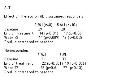
The aim of this study is to assess the efficacy and safety of 2 different doses of alfa-2b interferon combined with ribavirin in HCV+ patients with normal ALT. To qualify for this study, patients had to have a biopsy consistent with chronic hepatitis; normal ALT on 2 or more occasions at least 3 months apart; compensated liver disease. Patients were randomized to receive one of two regimens: group 1- IFN alf-2b 3Million Units 3 times per week + ribavirin 1000-1200 mg/day; or, group 2- interferon alfa-2b 5 MU 3 times per week + ribavirin 1000-1200 mg/day. Treatment was for 48 weeks, and if patients were HCV-RNA positive at week 24 they could stop therapy. There was a follow--up period of 24 weeks following therapy cessation to assess the 72 week SVR.
There were 28 patients in each group, 41% male; 69% genotype 1 in 3 MU group and 81% genotype 1 in 5 MU group. 24% genotype 2/3 in 3 MU group vs 11% in 5 MU group. Mean ALT was 35 in group 1 and 31 in group 2. Fibrosis stage 0-1: 18% in each group. Fibrosis stage 2-4 11% in group 1, 9% in group 2.
16 patients had stage 0. 20 patients had stage 1. 13 patients had stage 2. 6 patients had stage 3. 1 patient had stage 4. So, 7/28 (25%) patients with apparently normal ALT had stage 3/4.
RESULTS
The overall response rate was 32% for all patients , but 28% for the 3 MU group and 40% in the 5 MU group. Genotype 1 patients receiving 5 MU had 40% SVR vs 10% for patients receiving 3 MU. For genotype 2/3 patients over 80% receiving 3 MU had SVR and 70% receiving 5 MU had SVR.
3 patients relapsed after ending 48 weeks treatment. 4 patients had viral breakthrough on therapy. 32% of patients with stage 0,1 (n=36) had SVR and 30% with stage 2, 3, 4 had SVR. 40% of patients with <2 million HCV-RNA had SVR and 20% with >2 million had SVR.
|
|
 |
| |
Adverse Events
5 patients experienced ALT elevations. All were reportedly transient, one with transient 2.2 bilirubin. They were not more than 2x upper limit of normal. There were 2 serious adverse events: 1 suicidal ideation; confusion, anxiety. 9/56 patients (16%) discontinued due to an adverse event. Dose modifications were similar in both groups. There were no unexpected adverse events.
Jacobson concluded there was a trend towards a better response with IFN 5 MU in patients with genotype 1. ALT elevations were infrequent, mild, and transient. Response in stage 2-4 was similar to stage 0-1. ALT was reduced during treatment and in SR. AE, and serious AE was similar as in other studies. Response rates were comparable to patients with normal ALT. The trend towards superiority of IFN 5 MU leaves open question on optimal dosing. Combination therapy is safe- de novo ALT elevations are seldom problematic. Treatment should be individualized based on patient characteristics, including history. Therapy should not be ruled out based on ALT alone. This trial was conducted before pegylated IFN was available, so studies using peg IFN in patients with normal ALT are in progress.
|
|
| |
| |
|
|
|