 |
|
|
| |
Mitochondrial function, morphology, and metabolic parameters improve after switching from a stavudine- to a tenofovir-containing regimen:
SNAP (Switch Nucleoside Analogues Protocol)
|
| |
| |
Reported by Jules Levin
8th Intl Workshop on Adverse Drug Reactions and Lipodystrophy in HIV
Sept 2006, San Francisco
Courtney Kim1, Robert Murphy2, Baiba Berzins2, Jill Weinstein2, Jessica Shore2, Barbara Da Silva3, Elizabeth Belsey4, and Mariana Gerschenson1
1University of Hawaii, 2Northwestern University, 3Abbott Laboratories, 4SigmaClinical
ABSTRACT
Background: The pathogenesis of lipoatrophy in HIV-infected patients has been
associated with mitochondrial changes in fat tissue associated with NRTI therapy. Substitution by the nucleotide analogue tenofovir may result in improvement in mitochondrial function, morphology, and metabolic parameters.
Methods: Patients from Protocol M97-720 (n=10) receiving stavudine (d4T), lamivudine (3TC), and lopinavir/ ritonavir (Kaletra®) for 7 yrs were switched from
d4T to tenofovir (TDF) and followed for 48 wks. Subcutaneous fat tissue biopsies, fasting metabolic tests, HIV RNA and CD4 assays, and DEXA scans of fat content were obtained at baseline and 48 wks. MtDNA copies/cell and fat tissue morphology was examined by real-time PCR and transmission electron microscopy (TEM).
Results:
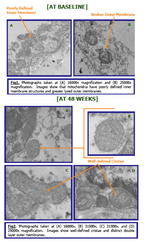
At baseline: TEM showed 1-2 mitochondria/preadipocyte and 1
mitochondria/adipocyte. Mitochondrial cristae (inner membrane) were lacking or
poorly defined.
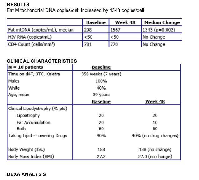
Median fat mtDNA=208 copies/cell. Median HIV RNA < 50 copies/ml, median CD4 count 781cells/mm3, on examination 2 patients had lipoatrophy, 2 fat accumulation, and 6 both; 4/10 were receiving lipid-lowering drugs.
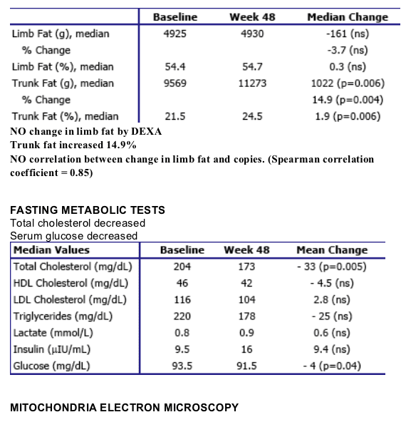
Median limb fat was 4925 and trunk fat 9569 g by DEXA, total, HDL, and LDL cholesterol were 204, 46 and 116 mg/dL, respectively, triglycerides 220 mg/dL, glucose 93.5 mg/dl, lactate 0.8 mmol/l, and insulin 9.5 uIU/ml.
48 weeks after switching to TDF: TEM showed an increase in platelets and endothelial cells suggesting increased vascularization, increased collagen in the adipocytes, and less preadipocytes. Mitochondrial numbers were increased 100%. The mitochondrial inner membranes were intact and contained well-defined cristae. The median fat mtDNA increased by 1343 copies/cell (p=0.002). There was no change in limb fat by DEXA, however, trunk fat increased
14.9 % (1022 grams; p=0.006). (note: This study was designed to be an intense mitochondrial study and may be too small to see limb fat changes).
Fasting total cholesterol decreased 33 mg/dL (p=0.04) and serum glucose decreased 4 mg/dl (p=0.005). No significant change in the other measured parameters including HIV RNA and CD4.
Conclusions: Fat mitochondrial function and morphology improved in adipose
tissue of patients switched from d4T to TDF while remaining on Kaletra. Total
cholesterol and glucose were decreased and trunk fat increased. This study suggests that switching from d4T to TDF results in improvement in fat mitochondria.
BACKGROUND
The pathogenesis of lipoatrophy in HIV-infected patients has been associated with mitochondrial changes in fat tissue associated with nucleoside reverse transcriptase inhibitor (NRTI) therapy. Substitution by the nucleotide analogue tenofovir (TDF) may result in improvement in mitochondrial function, morphology, and metabolic parameters.
METHODS
Patients enrolled in the Abbott M97-720 protocol at Northwestern University who
were receiving a stavudine (d4T), lamivudine (3TC), and lopinavir/ ritonavir
(Kaletra®) regimen for at least 6 years were switched from d4T to tenofovir (TDF).
1. Patients (N = 10) who consented to participate in the SNAP study had the following evaluations performed before switching to tenofovir (baseline) and after 48 weeks on the tenofovir-containing regimen:
2. Subcutaneous adipose tissue biopsy (4-mm skin punch biopsy on proximal lateral thigh. Fat tissue frozen in RNAlater or stored in gluteraldehyde.)
3. Mitochondrial morphology in fat tissue was examined by transmission electron
microscopy (TEM). Tissues fixed in gluteraldehyde were then post-fixed with 1%
osmium tetroxide and subsequently dehydrated in a series of graded alcohol
washes. Following, tissues were embedded in an epoxy resin substrate and sliced into thin micrometer cross-sections using a ultramicrotome. Sections were
mounted on slot or mesh grids and images were photographed at 16000x,
25000x, and 32500x magnifications with a LEO 912EF transmission electron
microscope.
4. Mitochondrial function was assessed by quantifying mitochondrial DNA copies/cell (fat tissue) using Real-Time PCR analysis.
5. Clinical Evaluations:
-- Whole body DEXA scans of fat content
-- Fasting metabolic tests (total, HDL, and LDL cholesterol, triglycerides, glucose, insulin, lactate)
-- Plasma HIV-1 RNA levels, CD4 cell counts
-- Evaluation of lipodystrophy of arms, legs, and face by clinician
Nonparametric signed rank test was used to assess median changes from baseline to week 48. Spearman correlation coefficient was calculated for the changes in limb fat and changes in mtDNA.
|
| |
|
|
|
|
|