 |
|
|
| |
Characterization of Maraviroc Resistance in Patients Failing Treatment with CCR5-Tropic HIV-1 in MOTIVATE 1 and MOTIVATE 2s
|
| |
| |
Reported by Jules Levin
XVI International HIV Drug Resistance Workshop. Barbados, 12-16 June 2007
Julie Mori1, Michael Mosley1, Marilyn Lewis1, Paul Simpson1, Johnathan Toma2, Wei Huang2, Jeannette Whitcomb2, Giuseppe Ciaramella1 and Mike Westby1
1Pfizer Global Research and Development, Kent, UK 2Monogram Biosciences, California, USA
AUTHOR CONCLUSIONS
The preclinical findings of MVC resistance were predictive of the clinical findings.
Phenotypic markers of reduced MVC susceptibility were identified using the PhenoSense Entry assay for virus populations derived from plasma samples.
Plateaus in MPI (maximal percentage inhibition) were identified as a phenotypic marker of MVC resistance in virus from 4/12 patients following failure on blinded MVC and from one patient in the placebo arm who subsequently received open-label MVC.
These were confirmed by clonal analyses and susceptibility testing of replication-competent viruses in PBL assays.
Mutations in the V3 loop of gp120 were identified in viruses that showed a plateau in MPI <95%, but the pattern of amino acid changes was different between patients.
Multiple pathways to resistance present a challenge for the development of genotypic algorithms for MVC resistance.
Shifts in IC50 (without evidence of a plateau in MPI) were not identified as a common phenotypic marker of MVC resistance in patients failing therapy with CCR5-tropic HIV-1.
For one patient a approximate 3-fold shift in IC50 was seen; the phenotype was reproduced in PBL.
Eight of the 12 patients who failed treatment with MVC with an R5 virus showed no phenotypic marker of MVC resistance.
This suggests that the majority of patients failing with an R5 virus did not fail as a result of acquiring resistance to MVC.
Failure in these patients is likely to be associated with other factors.
ABSTRACT 10
Characterization of maraviroc resistance in patients failing treatment with CCR5-tropic virus in MOTIVATE 1 and MOTIVATE 2
J Mori1, M Mosley1, M Lewis1, P Simpson1, J Toma2, W Huang2, J Whitcomb2, G Ciaramella1 and M Westby1
1Pfizer Global Research and Development, Kent, UK
2Monogram Biosciences, California, USA
BACKGROUND: Maraviroc-resistant viruses generated in vitro appear able to use either maraviroc-bound CCR5 or free CCR5 as a coreceptor for cell entry. This mechanism of resistance is characterized phenotypically by dose-response curves with a reduced maximal percentage inhibition (MPI). To identify phenotypic and genotypic markers of maraviroc resistance, in vivo, samples from 37 patients in the Phase III clinical studies (MOTIVATE 1 and 2) who failed therapy with a CCR5-tropic virus were analysed.
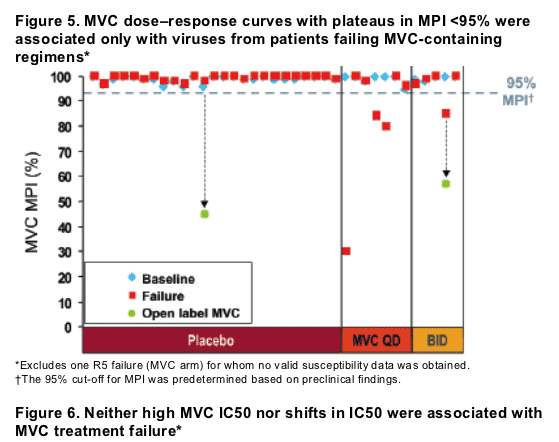
METHODS: Paired baseline and on-treatment samples were tested for maraviroc susceptibility using an Envelope (Env)-pseudotyped virus assay (PhenoSense Entry assay). Samples were identified for further investigation if one of three pre-defined criteria were met: plateau in MPI <95%, IC50 fold-change compared to reference virus (FC) outside the normal range (>1.95), or >two-fold change in IC50 between baseline and on-treatment samples. In these cases, susceptibility testing, gp160 sequencing and tropism confirmation were performed on 12 Env clones from each baseline and on-treatment sample. For five patients, Env genes were cloned into NL4-3, producing replicationcompetent viruses for testing in PBL.
RESULTS: No plateaus in MPI <95% were seen for any of 37 baseline viruses or for on-treatment virus from the 25 placebo patients. In contrast, plateaus in MPI <95% were seen for on-treatment samples from 4/12 patients following failure on blinded maraviroc and 2/3 patients subsequently followed onto open-label maraviroc. There was no significant difference in on-treatment maraviroc IC50
FCs between treatment groups (P=0.13, ANCOVA), although the on-treatment virus from one patient failing maraviroc demonstrated a small (threefold) shift from baseline. Clonal analysis confirmed results for the original population samples and results obtained in PBL were consistent with observations in the PhenoSense assay. Amino acid changes from baseline were identified in the V3
loops of samples yielding a plateau in MPI. Site-directed mutagenesis confirmed the importance of the V3 mutations in conferring the maraviroc phenotype. The
amino acid changes differed between patients, reflecting the heterogeneity in gp160 sequence. There was no evidence of cross-resistance with the fusion inhibitor enfuvirtide.
CONCLUSIONS: These results validate the use of a reduced MPI as a phenotypic marker of maraviroc resistance in vivo and identify V3 loop mutations as likely conferring the resistance phenotype.
BACKGROUND
Maraviroc (MVC) is a CCR5 antagonist in Phase 3 clinical development for the treatment of HIV-1 infection. Viral escape from MVC will be different from resistance to current antiretrovirals (ARVs) because MVC:
--binds to a host protein rather than a viral protein
--is only active against CCR5-tropic (R5) virus
--is not a competitive inhibitor.1,2
In preclinical in vitro studies, MVC resistance:
--was not associated with a change in co-receptor tropism (except where this also occurred in control virus passaged in parallel without MVC)
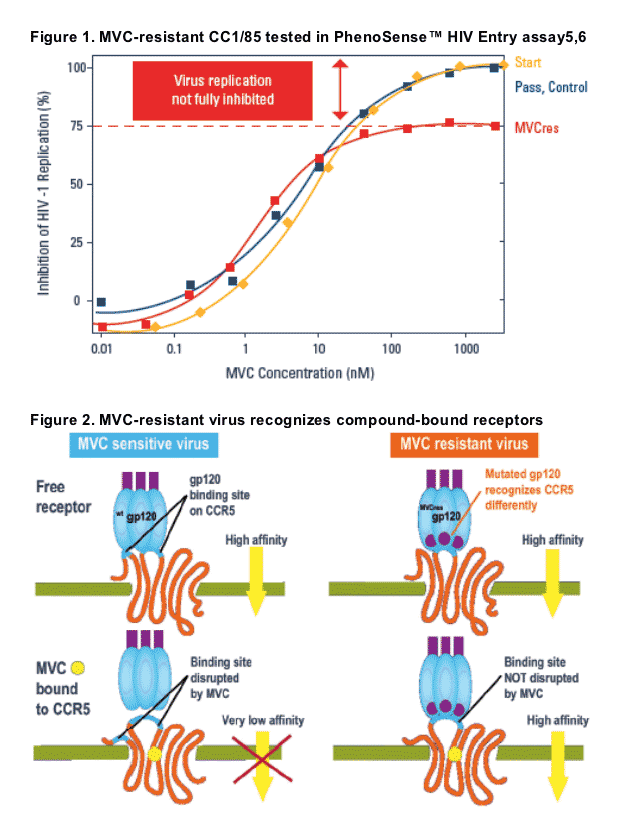
--was characterized by dose-response curves with plateaus in maximal percentage inhibition (MPI) rather than shifts in IC50 (Figure 1).
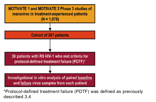
--was associated with amino acid changes in the V3 loop of the HIV-1 envelope glycoprotein (gp120)
--was NOT associated with cross-resistance to other investigational CCR5 antagonists or enfuvirtide (a HIV fusion inhibitor), efavirenz (a non-nucleoside reverse transcriptase inhibitor), and saquinavir (a protease inhibitor).
These findings are consistent with the use by MVC-resistant variants of both MVC-occupied and free CCR5 molecules to infect target cells (Figure 2).2
OBJECTIVES
To identify possible phenotypic and genotypic markers associated with MVC resistance in vivo by analyzing virus samples from 38 patients enrolled in the MVC clinical studies MOTIVATE 13 and MOTIVATE 24, who failed therapy with a CCR5-tropic (R5) virus.
To confirm the role of mutations in the V3 loop of gp120 in conferring an MVC-resistant phenotype using site-directed mutagenesis (SDM) of patient-derived viruses.
METHODS
MOTIVATE 1 and 2 are ongoing, randomized, double-blind Phase 3 trials in antiretroviral (ARV)-experienced patients with only R5 HIV-1 detected at screening (Trofile assay, Monogram Biosciences). MOTIVATE 1 was conducted in study centers across North America; MOTIVATE 2 was conducted in study centers across Europe, Australia, and North America.
Patients in both trials were randomized to receive placebo or MVC QD or BID (1:2:2). All three treatment groups also received an optimized background therapy (OBT) of 3-6 ARVs (pharmacokinetic boosting doses of retrovir not counted as an ARV).
The duration of both trials is 48 weeks with a planned interim analysis after 24 weeks.
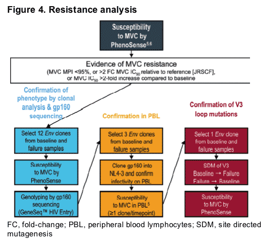
The origins of the patients included in this investigation of tropism changes are detailed in Figure 3.
Figure 3. Study design
This analysis included 38 patients with R5 HIV-1 who met criteria for protocol-defined treatment failure (PDTF)* taken from a cohort of 267 patients from MOTIVATE 1 & 2 phase 3 studies of maraviroc in treatment-experienced patients (n=1,076). The 38 patients received an investigational in vitro analysis of paired baseline and failure virus samples from each patient.
All analyses were conducted on blinded samples (except for the four open-label samples).
The Day 1 (pre-dose) plasma sample was used as the 'pre-treatment' baseline sample.
The last 'on-treatment' sample (following PDTF) was used as the failure sample.
Additional plasma samples from two patients during open-label MVC treatment were also tested.
Following unblinding of the study, it is now known that 25 patients received placebo and 13 received MVC either once- or twice daily.
RESULTS
Susceptibility to MVC by PhenoSense
No plateaus in MPI <95% were seen for any pre-treatment viruses or viruses from patients receiving blinded placebo-containing therapy (Figure 5).
Plateaus in MPI <95% were seen for on-treatment viruses from four patients failing blinded MVC therapy and from one patient in the placebo arm who subsequently received open-label MVC (Figure 5).
In general there was no increase in MVC IC50 from baseline for on-treatment viruses from patients failing blinded MVC therapy (Figure 6).
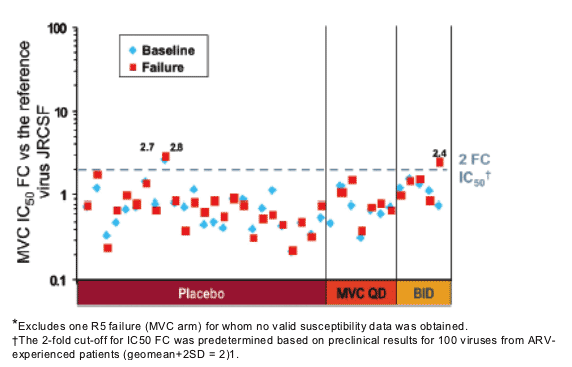
There was no significant difference in on-treatment MVC IC50 FC between treatment groups (P = 0.13, analysis of covariance).
-- Two patients showed an IC50 FC slightly higher than cut-off (Figure 6)
-- One patient received placebo
-- One patient received MVC twice daily and the increased IC50 was observed for the failure time point only.
Clonal analysis
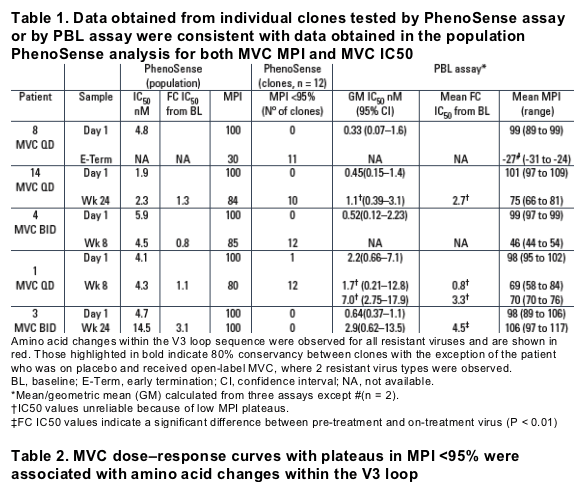
Data obtained from individual clones were consistent with data obtained in the population analysis for both MVC MPI and MVC IC50.
Data obtained using replication-competent NL4-3 in PBL assays were consistent with the results of the PhenoSense assay (Table 1).
A difference in plateau height was observed between the PhenoSense and PBL assays but the phenotype remained the same.
Genotypic analysis
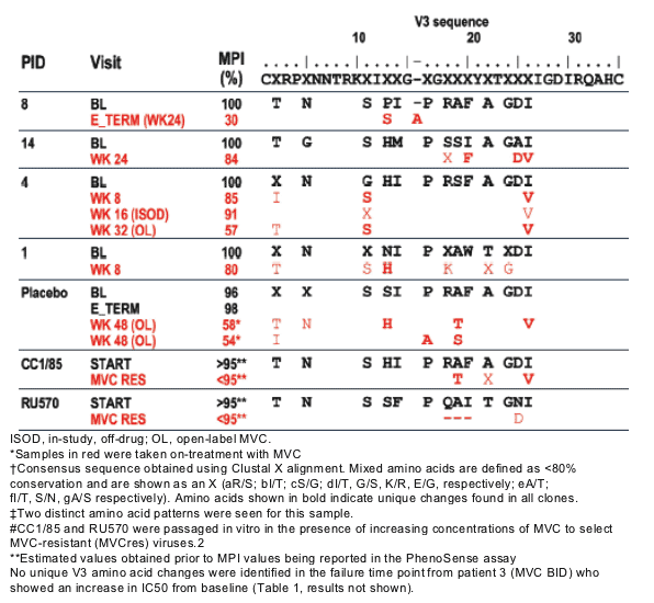
Genotypic analysis of the MVC-resistant virus revealed amino acid changes within the V3 loop (Table 2).
The amino acid changes within the V3 loop were different between patients' viruses.
Mutations at positions 13 or 26 in the V3 loop were observed in five patients.
Site-directed mutagenesis indicated the importance of these mutations in conferring the resistant genotype (Table 3).
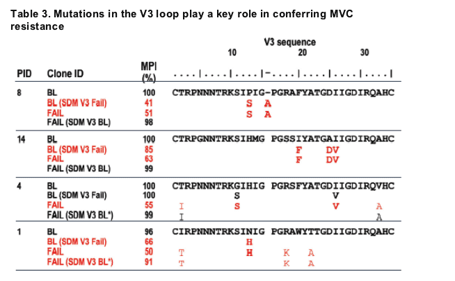
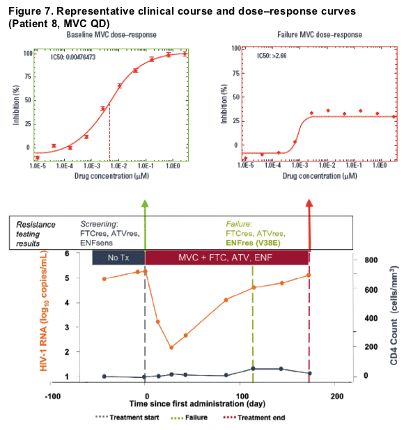
Patient 8:
-- 13S+16Ains were sufficient to cause MVC resistance in BL virus
-- 13S+16Ains were necessary for MVC resistance in the FAIL virus
-- Reversion to 13P+16Adel did confer MVC sensitivity in the FAIL virus.
Patient 14:
-- 20F+25D+26V were sufficient to cause MVC resistance in BL virus
-- 20F+25D+26V were necessary for MVC resistance in the FAIL virus
-- Reversion to 20I+ 25A+26I did confer MVC sensitivity in the FAIL virus.
Patient 4:
-- 11S+26V was not sufficient to confer MVC resistance in BL virus
-- 11S+26V were necessary for MVC resistance in the FAIL virus
-- Reversion to 11G+26I did confer MVC sensitivity in the FAIL virus.
Patient 1:
-- 13H was sufficient to confer MVC resistance in BL virus
-- 13H was necessary for MVC resistance in the FAIL virus
-- Reversion to 13N partially restored MVC sensitivity in the FAIL virus.
References
1. Dorr P, et al. Antimicrob Agents Chemother 2005; 49:4721-4732.
2. Westby M, et al. J Virol 2007; 81:2359-2371.
3. Lalezari J, et al. 14th CROI 2007. Presentation 104bLB.
4. Nelson M, et al. 14th CROI 2007. Presentation 104aLB.
5. Westby M, et al. J Virol 2006; 80:4909-4920.
6. Coakley E, et al. Curr Opin Infect Dis 2005; 18:9-15.
|
| |
|
|
|
|
|