 |
|
|
| |
Association between Peripheral Lipoatrophy and Bone Demineralisation in Treated HIV-positive Males
|
| |
| |
Reported by Jules Levin
10th International Workshop on Adverse Drug Reactions and Lipodystrophy in HIV, November 6-8 2008
Julian Falutz MD1, Esteban Martinez MD2, Waldo Belloso MD3, Leonard Rosenthall MD1
1McGill University Health Center*, Montreal, Canada 2Hospital Clinic, Barcelona, Spain 3Hospital Italiano, Buenos Aires, Argentina
AUTHOR SUMMARY & DISCUSSION
The rate of loss of mineralization at the hip region is relatively greater than at the lumbar spine in treated pts with decreased peripheral fat mass
There are significantly more pts with T-scores diagnostic of osteopenia or osteoporosis at the hip region vs the LS spine in pts who also have decreased peripheral fat mass
There is no difference in age, BMI, HIV-related response parameters, cigarette smoking, insulin resistance or concurrent use of PI-or NNRTI-HAART in pts with a lower vs higher proportion of peripheral to total body fat
Use of thymidine analogues may be associated with a relative preferential bone mineral loss at the hip region in pts with decreased peripheral fat mass
ABSTRACT
Introduction:
--Bone demineralization is increasingly recognized as an important component of the metabolic complications affecting HIV+ pts.
--In the general population multiple factors influence bone mineral density (BMD) including both regional lean and fat mass.
--In treated HIV pts body composition changes still include peripheral lipoatrophy, relative stability of lean mass and variable changes in central fat mass. We investigated factors associated with regional bone demineralization in HIV+ males.
Methods: A cohort of treated, clinically stable males who underwent DXA scans between 2001 and 2007 were characterized with regards to: 1-clinical (HAART usage, CD4s, HIV-VL, BMI, cigarette use, & serum testosterone levels); 2-metabolic (fasting lipid and glucose homeostasis); 3-DXA derived regional body composition (limb fat mass/total body fat mass ratio [%L/TBFM]); & 4-DXA derived BMD parameters (femoral neck [FN], total hip [TH] and lumbosacral [LS] spine t-scores). Differences between medians of variables were compared by the Mann-Whitney U test in pts grouped below and above the median value for the %L/TBFM. Differences between proportions were determined by Chi-square analysis.
Results:
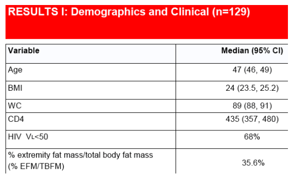
Data was available on 129 males (median age 47 (46-49), CD4's 438 (357-488), 68% with undetectable VL).
ARV use at the time of DXA acquisition revealed that 34% were on AZT, 40% on D4T, 13% on TDF, 64% on PI-HAART & 33% on NNRTI-HAART.
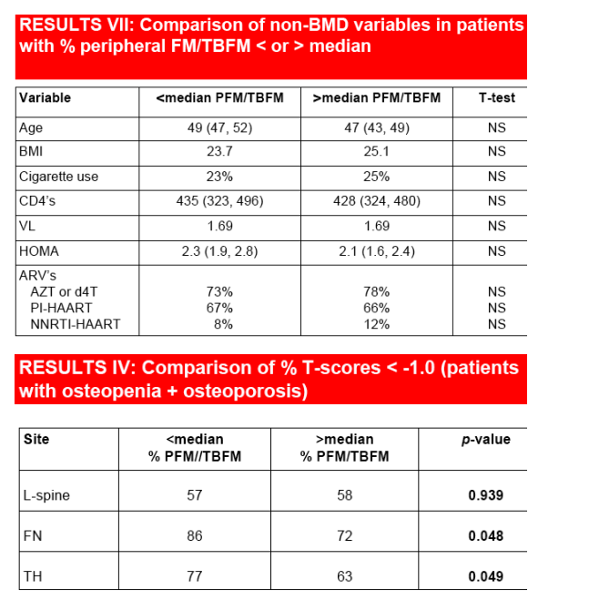
The median L/TBFM ratio was 36%. Median T score at the FN in pts with %L/TBFM <36% was -2.08(-2.4,-1.8) vs -1.84(-2.3,-1.2) in pts with %L/TBFM >36% (p=0.03).
Median T score at the TH in pts with %L/TBFM <36% was -1.81(-2.1,-1.4) vs -1.56(-2.0,-0.9) in pts with %L/TBFM >36% (p=0.11).
There was no difference in T scores at the LS spine in pts with %L/TBFM <36%>.
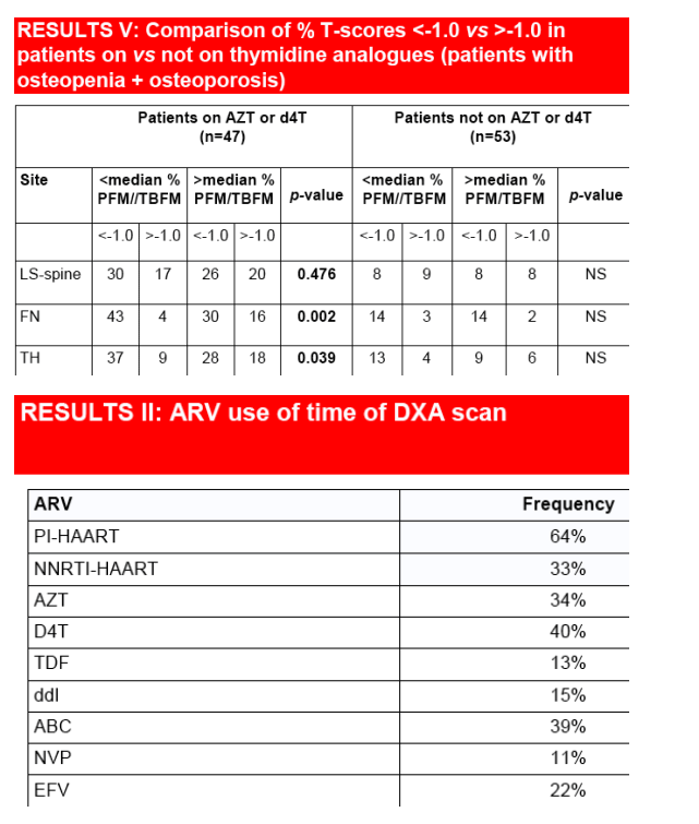
Comparing pts with %L/TBFM36%, there were more pts with osteopenia and osteoporosis at the FN (87% vs 71%, p=0.048), and at the TH (77% vs 62%, p=0.049), but not at the LS spine.
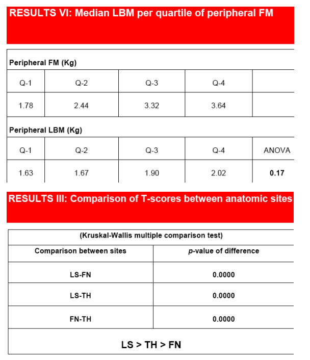
The limb lean mass did not change significantly for changes in limb fat mass.
There were no differences in pts with %L/TBFM 36% with regards to age, BMI, cigarette use, CD4's, VL, HOMA, or testosterone.
ARV use was similar in both groups except more pts with %L/TBFM<36% were on D4T, and more pts with %L/TBFM>36% were on AZT.
Conclusion:
--Progressive loss of peripheral fat mass, unrelated to loss of lean mass, is associated with decreasing BMD in the hip region but not at the LS spine.
--This LA associated bone demineralization may contribute to increasing hip related morbidity in treated patients, especially as they age.
--Both total body and regional BMD assessment should be considered and
efforts to prevent and treat peripheral LA instituted where possible.
INTRODUCTION
Premature bone demineralization is increasingly recognized in HIV(+) pts
The etiology of bone demineralization in HIV infection is multifactorial
Traditional non-HIV related factors contribute to bone demineralization in HIV infection
HIV-related determinants of bone demineralization include duration of HIV infection and possibly duration and type of antiretroviral drug exposure
Body composition is a determinant of bone mineral density (BMD) in control populations
In HIV negative populations bone demineralization occurs at different rates at the LS spine and at the hip region
Both lean body mass (LBM) and fat mass (FM) may contribute to BMD in control populations
In treated HIV infected pts, peripheral lipoatrophy [LA] occurs frequently
LBM remains relatively constant in clinically and immunologically stable treated pts
HYPOTHESIS
Peripheral lipoatrophy in pts with stable peripheral LBM is associated with regional bone demineralization
METHODS
·DXA scans (LUNAR) were done to determine BMD T-scores [WHO classifications] at 3 regions of interest: femoral neck (FN), total hip (TH) and lumbosacral spine (LS)
·DXA scans were used to determine regional body composition parameters
·A cohort of clinically stable, treated HIV(+) males who underwent routine DXA scanning between 2001-2007 was identified
·A retrospective chart review was performed to obtain clinical and metabolic parameters done within 3 months of the DXA scan
·Clinical and HIV-related data obtained included age, BMI, HIV viral load & CD4 counts, antiretroviral drug use at the time of DXA scanning, cigarette use, and HOMA
·Patients were divided into 2 groups: above and below the median of the % peripheral FM to total body fat mass ratio (% PFM/TBFM)
·The prevalence of T-scores Ġ-1.0 at the LS, FN & TH in the two groups were compared by the Chi-square test for proportions
·The prevalence of T-scores <-1.0 at the LS, FN & TH in pts on AZT or d4T vs those not on either NRTI was compared by the Chi-square test for proportions
·Differences between the clinical, HIV and metabolic variables within each group were compared by the Mann-Whitney U-test
·To determine change in LBM as function of FM in the lower limbs, the FM was stratified into quartiles and the median LBM of each FM quartile was compared by the Kruskal-Wallis one-way ANOVA on ranks
RESULTS
|
| |
|
|
|
|
|