 |
|
|
| |
GENDER AND GONADAL FUNCTION DIFFERENCES IN THE PREVALENCE OF BONE MASS REDUCTION: higher rates of osteopenia and osteoporosis for men vs women and at early ages, in their 40s.
|
| |
| |
Download the Poster PDF here
Reported by Jules Levin
CROI 2009 Feb 8-12 Montreal
Guaraldi G1, Zona S1, Vescini F2, Roverato A1, Orlando G1, Squillace N1, Garlassi E1, Luzi Kety1, Rochira V1, Lucia Zirilli1
1Metabolic clinic, Infectious Disease Department, University of Modena and Reggio Emilia, Italy - 2Endocrinologic clinic, University of Bologna, Italy
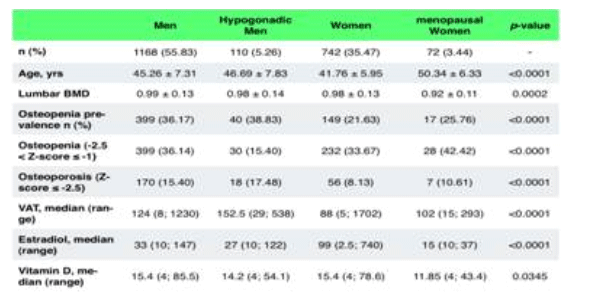
The table below shows osteopenia in 37% of men vs 23% in women, the difference is statistically significant. Osteoporosis prevalence was 16% in men vs 9% in women, also statistically significant. Of note, the average ages were 45-48 for men and 41-50 for women, very young to have so much prevalence of bone loss.
ABSTRACT
Background
To determine gender and gonadal function differences in the prevalence of Bone Mass Reduction (BMR or osteopenia) and to investigate its associated factors.
Methods
Cross-sectional survey in a cohort of 1962 consecutively recruited HIV-infected patients undergoing evaluation in a metabolic clinic. Male hypogonadism was defined for total testosterone <300 ng/mL. Female hypogonadism (menopausal) were defined for LH and FSH >20 ng/ml and 17 ß-estradiol<40 ng/mL. DXA scans of the lumbar spine and femoral neck were performed to determine z-score. For this analysis we excluded subjects using alendronate and/or VitD therapy, and primary hyperparathyroidism. BMR prevalence was defined for z-score<-1. Stepwise logistic regressions were performed to investigate independent predictors of BMR in men (both hypogonadal and eugonadal) and women in comparison to menopausal women. Endocrine, anthropometric, lifestyle, HIV and HAART variables were investigated for gender related differences.
Results
1207 (61.52%) males and 755 (38.48%) females were included in the analysis.
Both hypogonadal and eugonadal men categories were independent predictors of BMR in comparison to menopausal women (OR 3.59, CI: 1.94 - 6.65, p<0.001 and OR 3.43, CI: 2.19 - 5.37, respectively); cumulative PI exposure displayed a borderline risk (OR= 1.04, CI: 1.00-1.09, p=0.048). Protective factors were: 17 ß-estradiol (OR 0.96, CI: 0.94 - 0.98, p=0.003) and total lean/BMI ratio (OR per additional unit =0.39, CI: 0.24 - 0.61, p<0.001).
Conclusions
This large case series shows an unexpected high prevalence of trabecular bone mass reduction in men. We were unable to find an obvious explanation of this gender difference apart from a well known protective role of 17 ß-estradiol and lean mass. It's uncertain the clinical relevance of the borderline risk associated with PIs cumulative exposure.
We hypothesize it is necessary to move to a tissue level (a plausible difference of enzymatic activity of amortize has been previously suspected by our group) to understand the pathophysiological mechanism of gender difference
in BMR. This report underline that HIV related bone disease in men and women may be different, natural history of BMR according to gender is unknown.
AUTHOR CONCLUSIONS
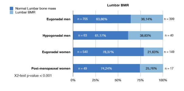
In order to interpret the higher prevalences of lumbar BMR in males we tried to explore any hormonal, anthropometric, viroimmunologic and therapeutic differences that may have had a different impact on trabecular bone in men vs. women. and in hypogonadic vs eugonadal population.
1. We confirm the expected lowest bone mass both in post-menopausal women and in hypogonadal men, stressing the obvious association between low levels of sex steroids and LBM. High prevalence of vitamin-D insufficiency was found both in maleas and in females but we were unable to find any difference in vitamin-D plasma concentration.
2. Unexpectedly total fat mass resulted to be a risk factor for LBM. We hypothesized that the extent of total fat mass may determine aromatase activity, necessary to produce 17-ß-estradiol and to concentrate it inside the bone micro involvement. The protective role of lean body mass on BMD has been describes by Dolan, as possibly associated with testosterone levels both in men and in women
3. Immuno-virologic parameters and HIV history were not different between groups. A borderline risk on BMR was associated with PIs cumulative exposure in our cohort.
Clinical relevance of this finding is uncertain.
This report underlines that HIV-related bone disease in men and in women may be different and natural history of LBM according to gender is unknown
We encourage a special attention on bone mass not only in hypogonadal men and women but in eugonadal men also.
BACKGROUND
In HIV infected patients receiving antiretroviral therapy (ART), reduced bone mineral
density (BMD) has been reported at increasing frequency
The guidelines of the International Society for Clinical Densitometry (ISCD) have
recommended to diagnose osteoporosis in men under 65 years and in fertile women on the basis of Z-score. The term "bone mass reduction" (BMR), identified for Z-score values below -1 SD, can better describe a homogeneous continuum of events leading to increased fracture risk and should substitute the terms "osteopenia" and "osteoporosis", particularly when a study clinical endpoint is based on BMD alone.
In HIV un-infected subjects, no differences are seen in the rate of bone mass loss until the onset of menopause, when women show a faster and more pronounced reduction of BMD than men due to lack of estrogens. We hypothesized that a gender difference in the prevalence and the natural history of bone disease may help to generate new pathogenic hypotheses in order to explain the high prevalence of BMR in HIV infected patients.
OBJECTIVE
The aim of our study was to determine both gender and gonadal function differences in the prevalence of BMR in HIV-infected patients and to investigate the risk factors associated with BMR.
METHODS
Cross-sectional observational study of consecutive HIV-infected patients recruited in an outpatient clinic (HIV Metabolic Clinic) at the University of Modena and Reggio Emilia, Italy, between January 2004 and June 2007.
Inclusion criteria were:
· Serologically documented HIV-1 infection
· Age >18 years
· Stable lipid-lowering and diabetes therapy for at least 6 months
Exclusion criteria were:
· Diseases affecting bone metabolism other than HIV infection
· Vitamin D and/or calcium supplements
· Therapy with bone active drugs (i.e. bisphosphonates, raloxifene, strontium ranelate, estrogens).
The whole cohort was divided in four groups:
1. Eugonadal men,
2. Hypogonadal men
3. Eugonadal women
4. Post-menopausal women
Male hypogonadism was defined for total testosterone <300 ng/mL. Menopause was defined for FSH >20 ng/ml and 17 ß-estradiol <40 ng/mL. Hypovitaminosis D was diagnosed for VitD plasma level < 30 ng/mL.
Femoral or lumbar BMR was defined by Z-score vauels lower that -1 (ISCD recommendation for epidemiological studies involving populations affected by chronic conditions leading to secondary osteoporosis).
Continuous normally-distributed variables were analyzed using ANOVA or T-test; non-normally-distributed variables using Kruskal-Wallis test or Mann-Whitney test. Differences in categorical variables were analyzed using the X2-test or the Fisher's exact test where appropriate.
Multivariable stepwise logistic regression models were performed to investigate independent predictors of lumbar BMR in men (both hypogonadal and eugonadal) and women in comparison to post-menopausal women. In order to analyze a single multivariable statistical model able to weight at the same time gender and hormone effect, we chose to generate the categorical variables "low testosterone" and "low 17-ß-estradiol", separately generated for men and women, using a cut off identified by the first lower quartile of hormones plasma level.
RESULTS
2092 DXA scan
130 pts excluded (4 hyperparatyroidisn, 126 undergoing treatment for bone disease
or Vit D supplementation)
1962 pts analysed
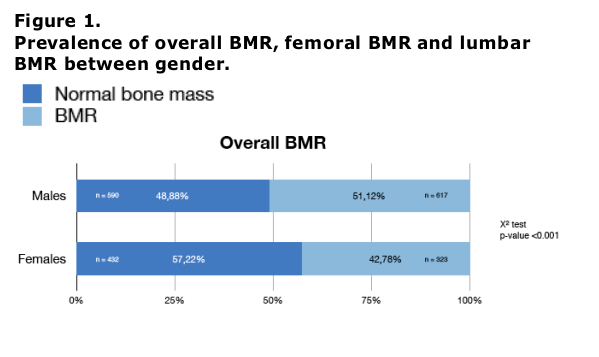
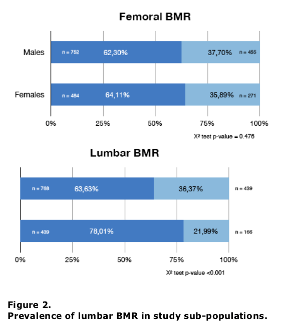
Prevalence of BMR, diagnosed at spine or hip level, was 47.91%, while the rate of
femoral BMR was 37.27% and that of lumbar BMR was 30.85%. Fig. 1 shows prevalence of BMR according to gender.
|
| |
|
|
|
|
|