 |
|
|
| |
Effects of Aging on HIV Pathogenesis: aging is associated with higher t-cell activation in women than men, effects of menopause on inflammation and immune activation...."
|
| |
| |
Click here to download the PDF
Reported by Jules Levin
CROI Feb 8-12, 2009, Montreal
Sumathi Sankaran-Walters1, Monica Macal1, Irina Grishina1, Mary K Miller2, Jason Flamm4, Thomas J. Prindiville3, and Satya Dandekar1
Departments of Medical Microbiology & Immunology1, Obstetrics and Gynecology2, and Internal Medicine3, University of California, Davis, CA, Kaiser Permanente Medical Group4, Sacramento, CA1 Pathogenesis
CONCLUSION BY AUTHORS:
"Aging is associated with higher levels of T cell activation in women
Effects of menopause on inflammation and immune activation plays an important role in HIV pathogenesis"
"HIV infection results in CD4+ T cell depletion in GALT
Progression is associated with T cell activation and tissue inflammation
Partial T cell restoration occurs with HAART
A threshold of CD4+ T cell restoration correlates with:
Reduced immune activation in GALT
Reduced inflammation in GALT
Suppression of viral replication"
Abstract
Background: Human Immunodeficiency Virus (HIV-1) is associated with loss of CD4+ T cells from the peripheral blood, over the course of infection. Gut associated lymphoid tissue (GALT) harbors the majority of CD4+ T lymphocytes in the body and is an important target for HIV. Severe depletion of intestinal CD4+ T cells occurs during primary HIV-1 infection and persists through the course of infection in the absence of anti-retroviral therapy. Inflammation and immune activation continue to be up regulated during Highly Active Anti-Retroviral Therapy (HAART) and are associated with lower levels of CD4+ T cell restoration. Studies suggest that younger HIV-1 infected patients have immune systems similar to uninfected older individuals characterized by a loss of regenerative capacity and an accumulation of aging T cells. However the effects of aging itself on HIV-1 pathogenesis are poorly understood. A growing proportion of HIV-1 infected individuals are now over 50 yrs old. In a chronic inflammatory setting, such as HIV infection, the pathogenic effects of HIV may be exacerbated by the altered hormonal levels during menopause.
Methods: Patient groups were analyzed in context of age (men) and hormonal status (women). We evaluated mucosal T lymphocyte subsets and activation using Flow cytometry and immunohistochemistry. Gene expression profiles and viral loads in adult HIV-1 positive patients of all age groups were analyzed using DNA microarray technology and real-time PCR.
Results: Immune activation was up regulated during HIV-1 infection and is associated with lower levels of CD4+ T cell restoration in all patient groups during HAART. Altered T cell subsets including central memory T cells and effector memory T cells were observed in post menopausal women. Increased levels of T cell activation were observed in post menopausal women and age matched men in both HIV positive and HIV-1 negative groups. Higher expression of pro-inflammatory factors was observed in CD4+ T cells from older negative controls. Similar results were observed in both HIV- 1 positive men and women in the older age group prior to and following the use of HAART.
Conclusions: Aging results in increased levels of inflammation as well as T cell activation in HIV-1 negative controls and these effects are exacerbated in the presence of HIV infection. The results of this pilot study will be extremely valuable in management of HIV disease worldwide.
Study Subjects
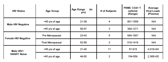
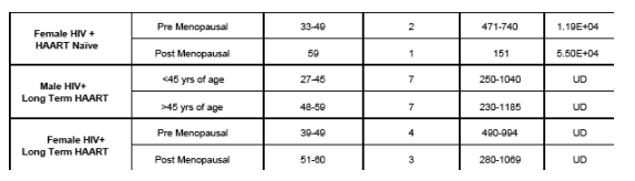
The acute phase of HIV infection occurs in the first few weeks to months following exposure. It is characterized by high viral loads in both peripheral blood and in GALT. The majority of CD4+ T cells are depleted in GALT during this stage of infection and T cell homeostasis is disrupted. All these changes result in the rapid onset of HIV associated enteropathogenesis. (HVL: High Viral Load.
LTNP: Long Term Non Progressor)
Increased T Cell Activation In Chronic HIV Infection
High levels of T cell activation is the hallmark of HIV infection. T cell activation is analyzed using the surface markers CD38 and HLA-DR which are expressed on activated T cells. Significantly higher levels of CD8+ T cell activation are observed in older men with HIV infection.
Long-term therapy could reduce, but not fully suppress, HIV RNA levels in the gut mucosa, though substantial gut CD 4 + T-cell restoration was still possible. These data also showed decreased levels of inflammation in GALT during therapy in patients with enhanced CD4 + T-cell restoration
In uninfected controls, women have higher levels of T cell activation in GALT compared to men. Furthermore, post menopausal women have higher levels of CD4+ T cell activation in peripheral blood as well as in GALT
Long term HAART is effective in suppressing viral replication in peripheral blood. However T cell activation in GALT persists. Compared to men, women have higher levels of CD4+ T cell activation in GALT. This could be due to a sexually dimorphic response as a result of their hormonal status.
Lower levels of Central Memory T cells were observed in GALT in both men and women of older age groups who were HIV negative or positive. These lower levels were maintained during the course of HAART in post menopausal women. This is in contrast to observations in peripheral blood.
Higher levels of FoxP3 positive CD4+ T cells were observed in GALT of post menopausal women who were HIV negative or positive and HAART naive. This effect was lost following HAART. These finding may be related to the level of the inflammatory response in GALT as seen using the transcriptional analysis shown below.
Increased transcription of several genes related to IL17 and inflammatory responses were up regulated in women compared to men in CD4+ T cells in peripheral blood
|
| |
|
|
|
|
|