 |
|
|
| |
HAART Interruption Increased Inflammation - IMPACT OF HAART INTERRUPTION ON PLASMA INFLAMMATORY
MARKERS ASSOCIATED WITH CARDIOVASCULAR DISEASE
FINAL 36-MONTH RESULTS FROM A RANDOMIZED STUDY
|
| |
| |
IAS Capetown July 2009
Reported by Jules Levin
M.M.Olmo*[1], C.Alonso-Villaverde[2], M. Penaranda[3], F. Gutierrez[4], J. Romeu[5], M. Larrousse[6], J. Curto[1], P. Domingo[7],
J.A. Oteo[8], P. Sanchez[1], D.Podzamczer[1], for the STOPAR STUDYgroup. Spain.
AUTHOR CONCLUSIONS:
HAART interruption lead to an increase in cardiovascular inflammatory markers after 36 months in comparison with continous therapy. As these cytokines correlate with endothelial damage and development of atherosclerotic plaque our data may partially explain the harmful cardiovascular effect of HAART-interruption.
THE HAART INTERRUPTION GROUP PRESENTED AN INCREASE IN SOME CARDIOVASCULAR INFLAMMATORY BIOMARKERS
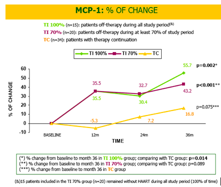
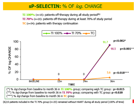
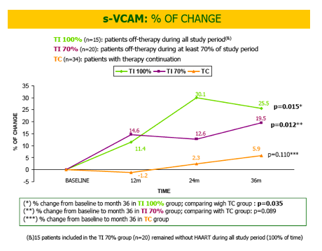
(MCP-1,sP-selectin, sV-CAM-1, sCD40L) IN COMPARISON WITH THE CONTINOUS THERAPY GROUP AFTER 36 MONTHS .
THESE CYTOKINES CORRELATE WITH PROGRESSIVE ENDOTHELIAL DAMAGE, DEVELOPMENT OF ATHEROSCLEROTIC PLAQUE AND EVEN PLAQUE DESTABILITAZION.
NEVERTHELESS, NO CARDIOVASCULAR EVENTS WERE OBSERVED IN ANY OF THE INTERRUPTION GROUP PATIENTS
OUR DATA MAY PARTIALLY EXPLAIN THE HARMFUL CARDIOVASCULAR EFFECT OF HAART INTERRUPTION AND CLAIM AGAINST THIS STRATEGY.
LONGER FOLLOW UP PERIODS AND FURTHER SUDIES ARE NEEDED TO DETERMINE THE PROPER BIOMARKERS FOR CARDIOVASCULAR DISEASE IN HIV-INFECTED PATIENTS
ABSTRACT
Background: HAART interruption has been associated with increased cardiovascular (CV) risk. We analyzed the influence of a CD4-guided HAART interruption on plasma inflammatory markers associated with CV disease.
Methods: Substudy of a randomized, multicenter, open, 36-month completed trial. HIV+ adults with viral supression >6 months and CD4 >500 (nadir >100) mainly NNRTI-treated, were allocated to therapy continuation (TC) or CD4-guided therapy interruption (TI). IL-6, IL-8, MCP-1, sVCAM-1, sCD40L, sP-selectin and t-PA were measured (multiplex cytometric bead-based assay (FlowCytomix Multiplex; BenderMedsystems; Austria))) at baseline, months 12, 24 and 36. TC-pacients and TI-patients with at least 70% time off-HAART were analyzed. Data presented as % of change or % of log change from baseline and median values; 36-month results.
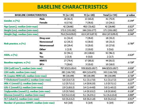
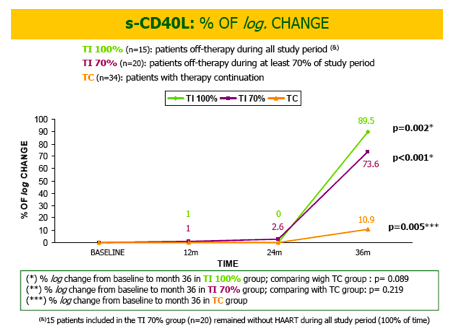
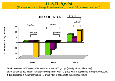
Results: We could analyze baseline and month 36 samples in 54 (34 TC, 20 TI) out of 77 (44 TC, 33 TI) included patients. All baseline characteristics were comparable: 77.9% were men, age 41, 36.4% homosexual, 32.5% former drug users, 6.5% AIDS and 83.1% NNRTI-regimen; total cholesterol/HDL-c/ LDL-c: 5.1/1.3/3 mmol/L, triglycerides 1.7 mmol/L, glucose 5.1 mmol/L. MCP-1, sP-selectin, sV-CAM-1 and sCD40L increased from baseline to month 36 in TI-patients (p=0.026, 0.020, 0.089, 0.219 respectively ) in comparison with CT-patients. When TI-pts 100% time off-HAART (instead of 70%) were analyzed (n=15),MCP-1 and sP-selectin results were confirmed, and, moreover, sV-CAM-1 reached a significant increase (p= 0.035), and sCD40L had a trend to increase (p=0.089). IL-6 decreased in TC-patients while remained stable in TI-patients but differences were not significant between groups. Not conclusive data were found in t-PA and IL-8.
Conclusions: HAART interruption lead to an increase in cardiovascular inflammatory markers after 36 months in comparison with continous therapy. As these cytokines correlate with endothelial damage and development of atherosclerotic plaque our data may partially explain the harmful cardiovascular effect of HAART-interruption.
BACKGROUND
HIV infection has been related to an increased level of pro-inflammatory markers (Joven J et al. Clin Chim Acta. 2006 Jun368 (1-2):114-9). Moreover, viral replication both in naïve patients (Ross AC et al. JAIDS 2008 Dec 15; 49(5):499-506)and in those interrupting HAART (Phillips A et al.Antivir The. 2008;13(2):177-87)has been associated with an increase in cardiovascular risk. Theoretically, the uncontrolled viral replication leads to an increase in several inflammatory proteins with effects on the endothelial wall and consequently on the development of the atheroma plaque. (Kuller et al.SMART Study.CROI 2008. Abstract 139; Calmy et al. STACATTO Study.CROI 2008. Abstract 140, Olmo et al. STOPAR Studymonth 24. CROI 2009. Abstract 116)
OBJECTIVE
To assess the influence of a CD4-guidedHAART interruption strategy on inflammatory biomarkers associated with cardiovascular disease
PATIENTS & METHODS
STOPARSTUDY: Randomized, open, multicenter, 36 month completed trial in adult HIV-infected patients on stable HAART with > 500 CD4 cells/mm3 for ≥6 months with viral supresssion for 3months . Patients were randomized to therapy continuation (TC) or to therapy interruption (TI). HAART was reinitiated if CD4<350cells/mm3 and rediscontinued if CD>500 with viral supression. Primary endpoints: disease progression, death, CD4 <200cells/mm3 or virological failure.
INFLAMMATORY MARKERS SUBSTUDY: Interleukin (IL)-6, IL-8, monocyte chemotactic protein (MCP-1), soluble vascular cell adhesion molecule (s-VCAM), soluble P-selectin (sP-selectin), tissue plasminogen activator (tPA) and soluble CD40 ligand (sCD40L) were measured at baseline, month 12, 24 and 36 using a multiplex cytometric bead-based assay (Human Cardiovascular 7plex FlowCytomix Multiplex; BenderMedsystems GmbH, Viena, Austria)and were analyzed on a EPICS-XL-MCL flow cytometer(Beckman Coulter; IZASA; Barcelona; Spain)following the manufacturer's protocol.
We could analyze baseline and month 36 samples in 54 (34 TC, 20 TI) patients. TI-patients with ≥70% and TI-patients with 100% (n=15/20) of time off-HAART were compared with CT-patients. MCP-1, s-VCAM and t-PA results are expressed as % of change from baseline. Logarithmic transformed values instead of absolute values of IL-6, IL-8, sP-selectin and sCD40L were used due to the skewed distribution shown by these variables, therefore their results are expressed as % of logarithmic change from baseline.
Statistical analysis: data are presented with median % of change from baseline and with median absolute values at each time point. Statistical tests used to compare between and within groups are shown under each table or graph, when appropriate. Final 36 monthdata are presented.
|
| |
|
|
|
|
|