 |
|
|
| |
Microbial Translocation and Hepatitis C Disease Progression among HIV-infected Women
|
| |
| |
Reported by Jules Levin
6th IAS Rome Italy July 17-20 2011
Audrey L French1,2, Denis M Agniel1, Charlesnika T Evans3, Mardge H Cohen1,2, Marion
Peters4 , Alan L Landay2 and Seema N Desai2 for the Women's Interagency HIV Study (WIHS)
1 CORE Center/Stroger (Cook County) Hospital. 2Rush University Medical Center, 3Northwestern University School of Medicine, Chicago IL, 4University of California, San Francisco, SF, CA, US
Abstract
Background: HIV/ HCV coinfection leads to more rapid liver disease progression (LDP) than HCV monoinfection. We sought to determine if gut microbial translocation (MT) and its inflammatory consequences contribute to LDP.
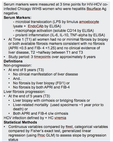
Methods: We compared markers of MT (lipopolysaccharide (LPS), endoCAb (IgM to LPS core), macrophage activation (soluble CD14 (sCD14)), and pro/anti-inflammation (IL-6, IL-10, TNF-α) measured by ELISA between HIV/HCV infected LDP and non-progressors (NP). Markers were measured at three timepoints (T). T1 for all women was a visit at which serum markers or liver biopsy were consistent with no/minimal fibrosis. T3 for NP was after a 4-6 year period during which biopsies or fibrosis markers did not change. Progression was defined as moderate/severe fibrosis by histology, imminent liver-related death or serum markers consistent with significant fibrosis at T3 4-6 years later. T2 was ~equidistant between T1 and T3. HCV infection was defined by HCviremia.
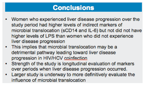
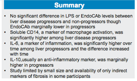
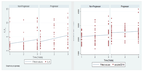
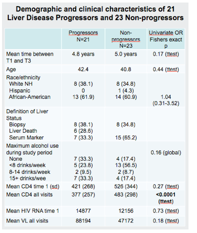
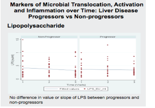
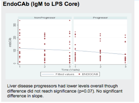
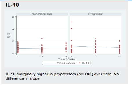
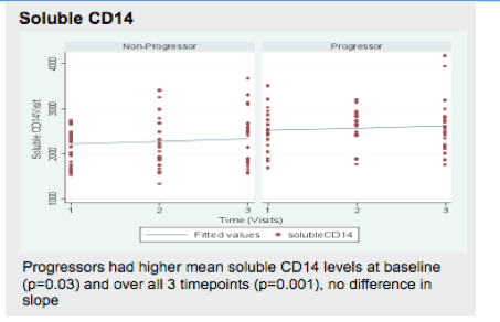
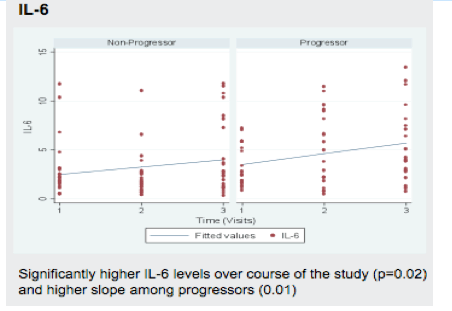
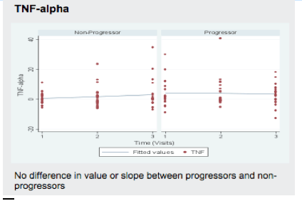
Results: Soluble markers were measured in 21 LDP and 23 NP. The median time between the T1 and 3 was 5.0 years. Median age, CD4 and HIVRNA for LDP and NP were 41.5 and 40.8 years; 410 and 426; and 14,110 and 12,156 respectively. LPS, endoCAb, IL-10, and TNF-α levels were not different in slope or quantity over time between LDP and NP. For IL-6 and sCD14 (graph), levels were higher overall in LDP (p=0.04 and 0.004, respectively) and, for IL-6, the slope was higher for progressors (p=0.03).
Conclusion: Markers of macrophage activation and inflammation were higher in HIV/HCV infected women during periods when liver disease progression occurred, however LPS did not differ between liver disease progressors and non-progressors.
Introduction
Early events in HIV infection deplete gut-associated lymphoid tissue leading to greater gut permeability.
There is a well-established link between microbial translocation and hepatic fibrosis in alcoholism, microbial translocation is also implicated in hepatic fibrosis with celiac sprue and graft vs host disease
There is a well-established link between HIV disease and hepatic fibrosis progression in HCV infection but the mechanism by which HIV leads to enhanced HCV disease progression is not established
Kuppfer cells, hepatic macrophages, are activated by a process involving bacterial cell wall components. Kuppfer cell activation leads to fibrosis through stellate cell activation.
We sought to determine if markers of microbial translocation and its downstream inflammatory consequences differed between HIV-HCV coinfected women who experienced more rapid liver disease progression and those who did not.
Methods
RESULTS
|
| |
|
|
|
|
|