 |
|
|
| |
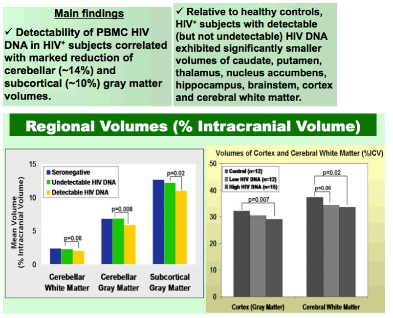
Detectable HIV DNA is Associated with Reduced Cerebellar and Subcortical Gray Matter Volumes
|
| |
| |
Reported by Jules Levin
CROI 2012
Kalpana J. Kallianpur1; Cecilia Shikuma1; Victor Valcour2; Bruce Shiramizu1; James Taylor1; Dominic Chow1; Gregory R. Kirk3; Beau K. Nakamoto1,4; Napapon Sailasuta5
1Hawaii Center for AIDS, University of Hawaii, Honolulu, HI; 2Memory and Aging Center, University of California at San Francisco, CA; 3Waisman Laboratory for Brain Imaging and Behavior, University of Wisconsin, Madison, WI; 4Straub Clinics and Hospital, Honolulu, HI; 5Huntington Medical Research Institutes, Pasadena, CA
ABSTRACT
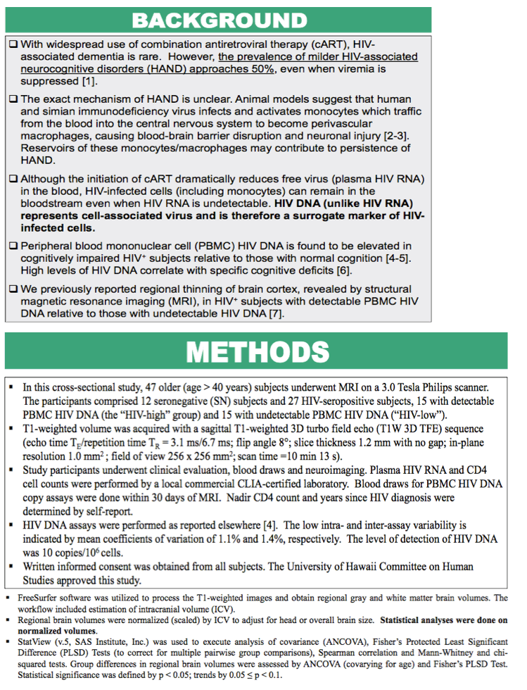
Background: HIV DNA is elevated in peripheral blood mononuclear cells (PBMC) of cognitively impaired HIV-seropositive patients despite highly active antiretroviral therapy (HAART). This cellular reservoir may reflect high levels of infected monocytes that trigger inflammatory processes upon crossing the blood-brain barrier. We assessed regional and global brain volumes as potential correlates of PBMC HIV DNA.
Methods: Cross-sectional analyses were conducted on T1-weighted 3-Tesla brain MRI data from participants in three groups: HIV+ subjects with undetectable PBMC HIV DNA, HIV+ with detectable HIV DNA, and seronegative (SN) controls. Segmentation with FreeSurfer yielded volumes of subcortical gray matter structures, brainstem, total cortical gray, cerebral white, and cerebellar gray and white matter. These were scaled by intracranial volume and compared among groups by ANCOVA controlling for age. Fisher's PLSD Test corrected for multiple pairwise group comparisons. Spearman correlation assessed relationships of brain volumes to clinical variables.
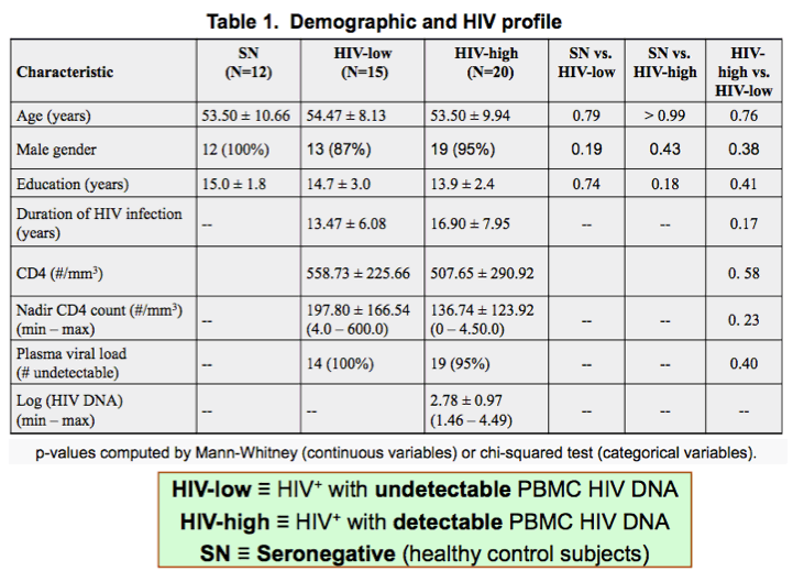
Results: We enrolled 47 subjects (44 male) with a median age of 54 years and 14 years of education. Fifteen had undetectable PBMC HIV DNA (<10 copies/106 cells), 20 had detectable HIV DNA (median=401, min=29, max=31,159 copies/106 cells), and 12 were SN. All HIV+ subjects were stable on HAART for ≥12 months, with plasma HIV RNA <50 copies/mL in all but one (158 copies/mL). The two HIV+ groups did not differ in age, education, duration of illness, current or nadir CD4 cell count.
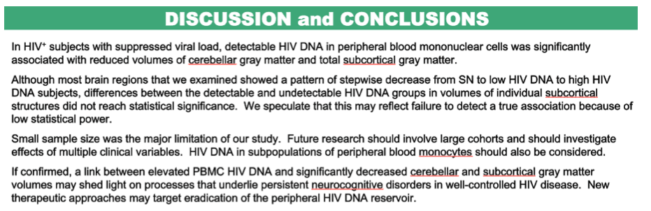
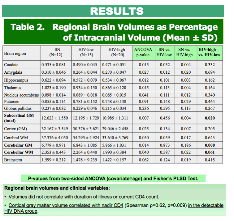
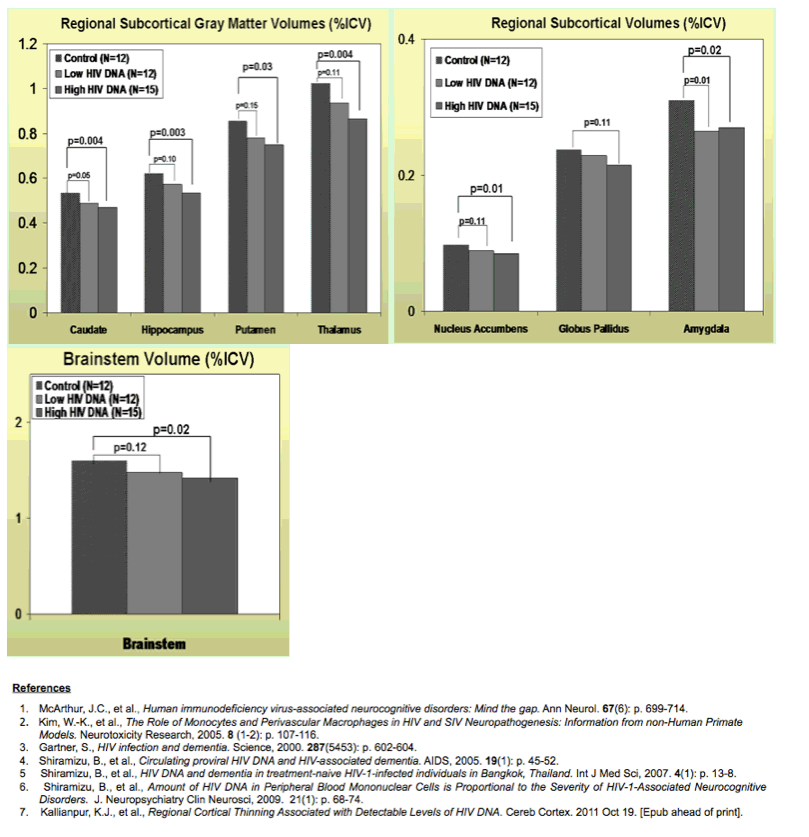
MRI revealed stepwise volumetric decrease of regional and total subcortical gray matter, cortex, brainstem, total cerebral white and cerebellar gray and white matter across SN, undetectable and detectable HIV DNA groups, with most reductions significant (p<0.05) between SN and detectable HIV DNA subjects only. Volumes did not correlate with duration of illness or current CD4 count. Cortical gray matter volume correlated with nadir CD4 in the detectable group (ρ=0.62, p=0.009). Relative to undetectable HIV DNA subjects, the detectable HIV DNA group exhibited significant 13% atrophy of total subcortical (p=0.02) and cerebellar (p=0.008) gray matter and a trend decrease in cerebellar white matter volume (-15%, p=0.06).
Conclusions: Severity of brain atrophy differs by PBMC HIV DNA detectability in well-controlled HIV disease and supports the role of incompletely treated peripheral reservoirs in the neuropathogenesis of cognitive impairment.
|
| |
|
|
|
|
|