| |
What Causes Kidney Decline in HIV+ / Factors associated with iohexol-based glomerular filtration rate slope over 36 months in HIV-negative and HIV-positive individuals
|
| |
| |
Download the PDF here
What causes GFR (kidney) decline in HIV+ ?
"Together, these findings suggest that uncontrolled HIV infection is associated with loss of kidney function and that durable viral suppression can normalize longitudinal GFR trajectory.....In conclusion, compared with a demographically and behaviorally similar sample of HIV-negative participants, we found HIV-positive participants had significantly lower iGFR at baseline, despite similar estimated GFR values in the two groups. In contrast, there was no significant difference in iGFR decline over time by HIV status. In the HIV-positive group, HCV coinfection was associated with a lower baseline iGFR and nonsuppressed viral load at baseline was associated with a more rapid subsequent decline in iGFR. Other findings support albuminuria and cardiovascular disease as being strongly linked to kidney function loss over time in HIV-positive persons.
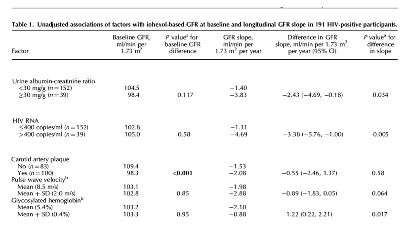
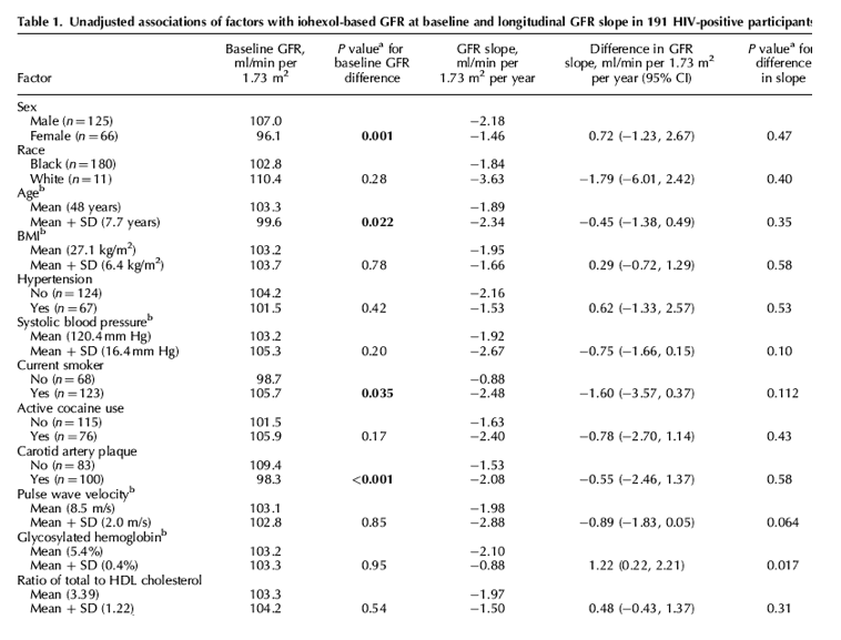
Among HIV-positive participants, factors that were statistically significantly associated with lower baseline iGFR included female sex, increased age, presence of carotid plaque, and HCV coinfection (Table 1). Tenofovir use and current smoking were significantly associated with higher iGFR at baseline.
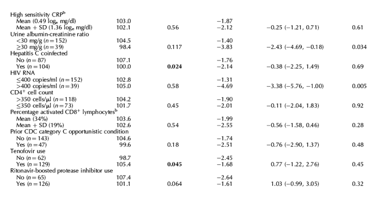
Only three factors were significantly associated with iGFR slope in HIV-positive participants. Higher glycosylated hemoglobin values [diabetes control] were associated with slower iGFR decline, whereas an albumin-creatinine ratio at least 30 mg/g was associated with a more rapid iGFR decline compared with those with lower levels of albuminuria (Table 1). Compared with patients with HIV RNA less than 400 copies/ml, those with nonsuppressed viral load had more rapid decline in iGFR (-1.31 vs. -4.69 ml/min per 1.73 m2 per year, P = 0.005). Higher pulse wave velocity values were borderline significantly associated with more rapid GFR decline. In a multivariable model containing variables that were significantly associated with iGFR slope in univariable models, nonsuppressed viral load (-3.09 ml/min per 1.73 m2 per year vs. suppressed viral load, 95% CI -5.48, -0.69) and glycosylated hemoglobin (1.24 ml/min per 1.73 m2 per year per 1 SD increase, 95% CI 0.26, 2.23) remained significantly associated with iGFR slope difference, whereas the association with albuminuria was nonsignificant (-1.98 ml/min per 1.73 m2 per year vs. no albuminuria, 95% CI -4.24, 0.28).
Glycosylated hemoglobin is also known as glycohemoglobin or as hemoglobin A1C (the main fraction of glycosylated hemoglobin). The level of glycosylated hemoglobin is increased in the red blood cells of persons with poorly controlled diabetes mellitus.. The normal level for glycosylated hemoglobin is less than 7%. Diabetics rarely achieve such levels, but tight control aims to come close to it. Levels above 9% show poor control, and levels above 12% show very poor control. It is commonly recommended that glycosylated hemoglobin be measured every 3 to 6 months in diabetes.
In HIV-positive patients we found adverse associations between viral factors (HCV coinfection and HIV RNA suppression), albuminuria, and cardiovascular disease indicators (carotid plaque and pulse wave velocity) and iGFR. We found no association between tenofovir or boosted protease inhibitor use and iGFR, with the exception of a significantly higher baseline iGFR in participants taking tenofovir compared with not taking tenofovir, an association that likely reflects selection bias.
In the HIV-positive group, we found HCV coinfection to be associated with a significantly lower iGFR at baseline, but no significant difference in iGFR slope over time. HCV has been associated with CKD and ESRD in other studies [13].....We also found associations between carotid plaque and lower baseline iGFR and between higher pulse wave velocity and more rapid iGFR decline.
These findings support the established link between cardiovascular and kidney disease in both the general [18] and HIV-positive populations [19]. In this nondiabetic sample, higher baseline glycosylated hemoglobin was associated with slower iGFR decline over time. The reason for this association is not clear, although diabetes has been associated with hyperfiltration [20,21] and our observed association may reflect increases in iGFR in patients with glucose intolerance at baseline.”
Pulse Wave Velocity (PWV) is a measure of arterial stiffness, or the rate at which pressure waves move down the vessel. It has been established as a highly reliable prognostic parameter for cardiovascular morbidity and mortality in a variety of adult populations including older adults, patients with end-stage renal disease, diabetes and hypertension. ...https://www.datasci.com/solutions/cardiovascular/pulse-wave-velocity.
Factors associated with iohexol-based glomerular filtration rate slope over 36 months in HIV-negative and HIV-positive individuals
AIDS Feb 16 2016
Lucas, Gregory M.; Atta, Mohamed G.; Zook, Katie; McFall, Allison M.; Mehta, Shruti H.; Fine, Derek M.; Stein, James H.; Schwartz, George J.
aDepartment of Medicine, Johns Hopkins University School of MedicinebDepartment of Epidemiology, Johns Hopkins University Bloomberg School of Public Health, Baltimore, MarylandcDepartment of Medicine, University of Wisconsin School of Medicine and Public Health, Madison WisconsindDepartment of Pediatrics, University of Rochester Medical Center, Rochester, New York, USA.
Abstract
Background: Monitoring kidney function is important in HIV-positive persons, but creatinine-based estimates of glomerular filtration rate (GFR) have limitations. There are little to no data available assessing GFR trends in HIV-positive persons using a gold-standard measure of GFR.
Methods: We measured GFR based on iohexol plasma disappearance (iGFR) annually for 3 years in nondiabetic, HIV-negative and HIV-positive volunteers with normal estimated kidney function. We used mixed linear models to evaluate factors associated with baseline iGFR and iGFR slope.
Results: One hundred HIV-negative and 191 HIV-positive, predominantly black individuals (median age 49 years) participated in the study and completed a total of 960 iGFR assessments over a median of 36 months. Despite similar estimated GFR at baseline, average iGFR values were lower in HIV-positive compared with HIV-negative participants (103.2 vs. 110.8, ml/min/1.73 m2, P = 0.004). However, subsequent iGFR slope was not significantly different in HIV-positive and HIV-negative participants. In the HIV-positive group, the presence of carotid plaque and hepatitis C virus coinfection were associated with significantly lower iGFR values at baseline. A nonsuppressed HIV RNA level at baseline was associated with a significantly more rapid iGFR decline compared with individuals with HIV RNA less than 400 copies/ml (-4.69 vs. -1.31 ml/min per 1.73 m2 per year, P = 0.005). Other factors significantly associated with iGFR slope included albuminuria and glycosylated hemoglobin.
Conclusion: Compared with HIV-negative persons, HIV-positive participants had significantly lower baseline iGFR, despite similar estimated GFR in the two groups. Nonsuppressed HIV RNA at baseline was associated with a more rapid iGFR decline over 3 years.
Introduction
The risks for acute kidney injury and chronic kidney disease (CKD) are increased in persons with HIV infection [1]. Intrinsic glomerular filtration rate (GFR) markers, most commonly serum creatinine, are widely used in clinical practice to monitor kidney function. However, creatinine is influenced by non-GFR factors and the accuracy of estimating equations can be poor [2]. Some studies suggest that estimating equations do not perform as well in HIV-positive compared with HIV-negative populations [3,4].
GFR measured with an exogenous filtration marker is considered the gold standard, but is time-consuming and rarely done in clinical practice. To date, several groups have presented cross-sectional analyses of exogenously measured GFR in HIV-positive participants [3-6]; however, few data are available regarding longitudinal GFR measures using a gold-standard method. Using GFR measurements based on iohexol plasma disappearance (iGFR), our objectives were to compare longitudinal iGFR trajectories in HIV-positive and HIV-negative participants, and assess the associations of baseline factors with baseline iGFR and iGFR slope in HIV-positive participants.
Methods
Study design
The Mr. Bean study is a cohort in Baltimore, Maryland to assess demographic, behavioral, and viral factors in the progression of kidney and cardiovascular disease [3]. Using flyers and local media, we recruited HIV-positive and HIV-negative participants in a 2 : 1 ratio, with the goal of achieving broadly similar demographic and behavioral characteristics in the two groups. Inclusion criteria included age 18 years or older and estimated GFR at least 60 ml/min per 1.73 m2. Exclusion criteria included history of radiocontrast allergy, pregnancy, diabetes mellitus, uncontrolled hypertension, or life threatening comorbidity.
Measurements and definitions
Participants completed a baseline study visit and up to three annual follow-up visits. At each visit, we collected demographic, behavioral, clinical, pharmaceutical, anthropomorphic, and laboratory data. Estimated GFR was calculated with the creatinine-based CKD-EPI equation [7]. Active cocaine use was defined by self-report or toxicologic detection of cocaine. Hepatitis C virus (HCV) infection was defined by antibody reactivity or detectable HCV RNA. We used flow cytometry on fresh whole blood to measure the percentage of CD8+ lymphocytes expressing an activated phenotype, defined by the coexpression of CD38+ and HLA-DR+ surface markers.
We measured iGFR at each visit according to a two-compartment model based on the disappearance of iohexol from serum, as described previously [3,8]. In addition, we measured pulse wave velocity, an index of vascular stiffness, by applanation tonometry and waveform capture at the carotid and femoral arterial sites using the SphygmoCor CPV System (West Ryde, Australia). At the baseline visit, trained technicians measured carotid intima-media thickness using Toshiba Aplio Ultrasound System. The presence of plaque, defined as a focal area of intima-media thickening at least 1.5 mm or 50% thicker than the neighboring wall, was assessed in the common and internal carotid arteries bilaterally [9].
Statistical analysis
We used Fisher's exact test and the Wilcoxon rank-sum test to compare categorical and continuous variables, respectively. We used separate linear regression models to calculate annualized iGFR slopes for each participant with at least two iGFR measures, and compared the distributions of the slopes in HIV-negative and HIV-positive participants. Next, to assess the association of factors with baseline iGFR and subsequent iGFR slope, we used multilevel mixed-effects linear regression models, with random intercepts for each individual and an exchangeable correlation structure for residuals, to account for repeated measures within individuals. Interaction terms between the variable of interest and time (in years) were included in regression models, such that separate coefficients represented the association between the factor and baseline iGFR and the association between the factor and the difference in iGFR slope, respectively. We used REDCap [10] for data management and Stata, version 13 (StataCorp, College Station, Texas, USA) for statistical analyses.
Ethical oversight
Participants provided written informed consent and the Johns Hopkins Medicine Institutional Review Board approved the study.
Results
A total of 291 participants (100 HIV-negative and 191 HIV-positive) had at least one valid iGFR and were included in the analysis. Of these, 26 (8.9%) had one, 30 (10.3%) had two, 66 (22.7%) had three, and 169 (58.1%) had four iGFR measures at annual visits, for a total of 960 iGFR measurements. The median (range) follow-up time was 36.4 months (0, 47.4).
Baseline characteristics have been presented previously [3].
[In conclusion, we found that CKD-EPI equations based on creatinine and cystatin C tended to be less accurate in HIV-positive than HIV-negative subjects. eGFRcys was significantly lessaccurate and more biased than both the widely-used eGFRcr equation and the combined eGFRcr-cys equation in HIV-positive individuals. Moreover, eGFRcys performance was strongly affected by uncontrolled HIV disease and T-cell activation indices. In contrast, eGFRcr performance was not modified by HIV-related factors or T-cell activation.]
The study sample was over 90% black and had a median age of 49 years (Table S1, supplemental digital content http://links.lww.com/QAD/A821). Compared with HIV-negative participants, HIV-positive participants were significantly more likely to be women (19 vs. 35%, P = 0.006), have a urine albumin-creatinine ratio at least 30 mg/g (6 vs. 20%, P = 0.001), and to have HCV (30 vs. 54%, P < 0.001). Estimated GFR values were similar in HIV-negative and HIV-positive participants (median 103 ml/min per 1.73 m2 in each group, P = 0.79). Ninety-one percent of HIV-positive participants were taking antiretroviral therapy, the median CD4+ cell count was 466 cells/μl, and 80% had HIV RNA below 400 copies/ml. There were no systematic differences in key baseline characteristics according to number of visits completed (Table S2, supplemental digital content http://links.lww.com/QAD/A821).
Among 265 participants (91 HIV-negative and 174 HIV-positive) that had 2 or more iGFR measurements, the distributions of individually calculated slopes were similar in HIV-negative and HIV-positive participants (Fig. 1a). Nine (10%) and 17 (10%) of HIV-negative and HIV-positive participants, respectively, experienced a more than 10 ml/min per 1.73 m2 per year decline in iGFR (P = 1.00). In a mixed-effects model, baseline iGFR was significantly lower in HIV-positive than HIV-negative patients (103.2 vs. 110.8, ml/min per 1.73 m2, P = 0.004); however, iGFR decline was not significantly different in the two groups [iGFR slope, -1.94 vs. -3.28 ml/min per 1.73 m2 per year in HIV-positive and HIV-negative participants, respectively; difference in slopes, 1.34 ml/min per 1.73 m2 per year, 95% confidence interval (CI), -0.22, 2.91, P = 0.092] (Fig. 1b).
Among HIV-positive participants, factors that were statistically significantly associated with lower baseline iGFR included female sex, increased age, presence of carotid plaque, and HCV coinfection (Table 1). Tenofovir use and current smoking were significantly associated with higher iGFR at baseline.
Only three factors were significantly associated with iGFR slope in HIV-positive participants. Higher glycosylated hemoglobin values were associated with slower iGFR decline, whereas an albumin-creatinine ratio at least 30 mg/g was associated with a more rapid iGFR decline compared with those with lower levels of albuminuria (Table 1).
Compared with patients with HIV RNA less than 400 copies/ml, those with nonsuppressed viral load had more rapid decline in iGFR (-1.31 vs. -4.69 ml/min per 1.73 m2 per year, P = 0.005). Higher pulse wave velocity values were borderline significantly associated with more rapid GFR decline. In a multivariable model containing variables that were significantly associated with iGFR slope in univariable models, nonsuppressed viral load (-3.09 ml/min per 1.73 m2 per year vs. suppressed viral load, 95% CI -5.48, -0.69) and glycosylated hemoglobin (1.24 ml/min per 1.73 m2 per year per 1 SD increase, 95% CI 0.26, 2.23) remained significantly associated with iGFR slope difference, whereas the association with albuminuria was nonsignificant (-1.98 ml/min per 1.73 m2 per year vs. no albuminuria, 95% CI -4.24, 0.28).
Discussion
In this longitudinal study of iGFR measurements in nondiabetic participants with clinically normal estimated kidney function at baseline, we found that baseline iGFR was significantly lower in HIV-positive than HIV-negative participants, despite nearly identical estimated GFR values in the two groups. However, HIV status was not significantly associated with subsequent iGFR slope.
In HIV-positive patients we found adverse associations between viral factors (HCV coinfection and HIV RNA suppression), albuminuria, and cardiovascular disease indicators (carotid plaque and pulse wave velocity) and iGFR. We found no association between tenofovir or boosted protease inhibitor use and iGFR, with the exception of a significantly higher baseline iGFR in participants taking tenofovir compared with not taking tenofovir, an association that likely reflects selection bias.
We found that HIV-positive participants had a lower baseline iGFR than a demographically similar HIV-negative comparison group, and that nonsuppression of HIV RNA was associated with a more rapid subsequent decline in iGFR in the HIV-positive group. Similarly, another study reported that viral suppression over time was associated with favorable changes in estimated GFR in patients with advanced HIV disease at baseline [11]. Additionally, a recent study from a North American consortium of HIV cohorts reported that the incidence of end-stage renal disease (ESRD) among HIV-positive patients that maintained durable viral suppression was similar to age-, sex-, and race-matched ESRD rates in the general population [12]. In contrast, we found no association between current CD4+ cell count and baseline iGFR or iGFR slope, consistent with a prior report [6]. Together, these findings suggest that uncontrolled HIV infection is associated with loss of kidney function and that durable viral suppression can normalize longitudinal GFR trajectory.
In the HIV-positive group, we found HCV coinfection to be associated with a significantly lower iGFR at baseline, but no significant difference in iGFR slope over time. HCV has been associated with CKD and ESRD in other studies [13]. However, it is difficult to disentangle HCV infection effects from associated socioeconomic and behavioral factors, including drug use [14-17]. In our study, we measured cocaine use, both by self-report and by urine drug screen. Despite a high prevalence of active cocaine use in the cohort, we found no association between cocaine use and baseline iGFR or iGFR slope.
We also found associations between carotid plaque and lower baseline iGFR and between higher pulse wave velocity and more rapid iGFR decline. These findings support the established link between cardiovascular and kidney disease in both the general [18] and HIV-positive populations [19]. In this nondiabetic sample, higher baseline glycosylated hemoglobin was associated with slower iGFR decline over time. The reason for this association is not clear, although diabetes has been associated with hyperfiltration [20,21] and our observed association may reflect increases in iGFR in patients with glucose intolerance at baseline.
This study is among the first to assess longitudinal kidney function changes in an HIV-positive sample using a gold-standard GFR measurement method. However, our study has limitations. First, the sample was predominantly African American, limiting the ability to extrapolate findings to other racial groups. Second, consistent with our research objectives, we excluded individuals with clinically decreased kidney function at baseline, preventing us from evaluating iGFR slopes in persons with overtly decreased kidney function. Third, a relatively small sample size and lack of longer-term follow-up may limit our ability to detect potentially clinically relevant associations, and multiple comparisons may increase the risk of identifying spurious associations.
In conclusion, compared with a demographically and behaviorally similar sample of HIV-negative participants, we found HIV-positive participants had significantly lower iGFR at baseline, despite similar estimated GFR values in the two groups. In contrast, there was no significant difference in iGFR decline over time by HIV status. In the HIV-positive group, HCV coinfection was associated with a lower baseline iGFR and nonsuppressed viral load at baseline was associated with a more rapid subsequent decline in iGFR. Other findings support albuminuria and cardiovascular disease as being strongly linked to kidney function loss over time in HIV-positive persons.
|
|
| |
| |
|
|
|