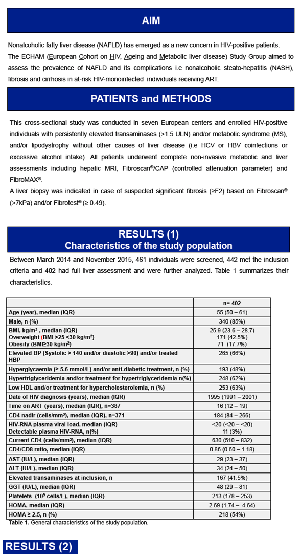
 |
|
|
| |
LIVER STEATOSIS AND FIBROSIS IN AT-RISK EUROPEAN HIV-MONOINFECTED PATIENTS - 64% with steatosis among those who had elevated LFTs and/or metabolic syndrome and/or lipodystrophy
|
| |
| |
Reported by Jules Levin
CROI 2017 Feb 14-16 Seattle WA
from Jules: this was a cross sectional study looking at HIV infected without hepatitis C or B but who had elevated liver enzymes - >1.5 >ULN which is not very high - they report 64% with significant staetosis & 9% with worse, NASH, and a many patients also with accompanying liver fibrosis. Many HIV+ individals have elevated liver enzymes but this appears to get past some of their doctors, not thinking perhaps they have fatty liver , the doctors may not be aware that elevated LFTs may be due to steatosis, they may think all HIV+ merely have this & they accept it without knowing what it could mean
Maud Lemoine1, Lambert Assoumou2, Patrick Ingiliz3, Pierre-Marie Girard4, Stephane De Wit5, Marc-Antoine Valantin2,6, Anja Huefner7, Jacqueline Capeau8, Dominique Costagliola2, Georg Behrens9.
1Imperial Coll London, London, UK,2Inst Pierre Louis d'Epidemiologie et de Sante Publique, Paris, France,3Cntr for Infectiology, Berlin, Germany,4Hopital Saint-Antoine, Paris, France,5Univ Libre de Bruxelles, Brussels, Belgium,6AP-HP, Paris, France,7Universitatsklinikum Hamburg, Hamburg, Germany,8Univ Pierre & Marie Curie, Paris, France,9Medizinische Hochschule Hannover, Hannover, Germany

Non alcoholic HIV-mono infected patients on ART with metabolic disorders are at high risk of liver steatosis, NASH and fibrosis. However, noninvasive markers of NASH and fibrosis by e.g. Fibroscan must be interpreted with caution in this population since we observed a poor accordance with histological results.
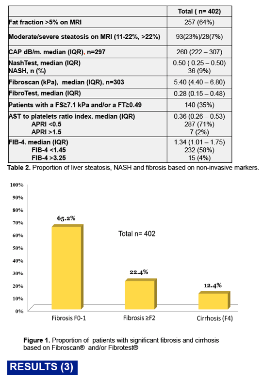
Proportion of patients with steatosis, NASH and fibrosis based on non-invasive markers
Hepatic MRI classified 257 (64%) patients with significant steatosis defined by a fat fraction (FF) >5%. Nash Test (Biopredictive) classified 36 (9%) patients with NASH. Usingnoninvasive markers of fibrosis, 140 (35%) had suspected significant liver fibrosis including 12.4% with cirrhosis (Table 2 and Figure1). However, the concordance between Fibroscan® and Fibrotest® for the diagnosis of fibrosis was poor (kappa coefficient 13%).
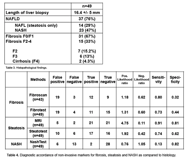
Of 140 patients eligible for liver biopsy, the procedure was performed in 50 (35%). Histological analysis was available for 49 patients and found NAFLD in 76% and NASH in 47%. Interestingly, significant fibrosis (F2/F3) and cirrhosis was confirmed in only 33% and 4% of patients, respectively (Table3). Using liver histology as a reference, the diagnostic performance of non-invasive markers to detect fibrosis, steatosis and NASH was poor (Table4). Only hepatic MRI showed a strong correlation with histologic steatosis (Positive likelihood ratio 4.75).
|
| |
|
|
|
|
|