 |
|
|
| |
NAFLD AASLD Guidelines - The diagnosis and management of nonalcoholic fatty liver disease: Practice guidance from the American Association for the Study of Liver Diseases
|
| |
| |
Download the PDF here
NATAP Fatty Liver / AASLD Fatty Liver - http://www.natap.org/liver.htm
AASLD: NAFLD Debrief AASLD 2017 - (11/29/17)
American Association for the Study of Liver Diseases AASLD
Washington DC
October 2017
from Jules: although HIV is not mentioned in these Guidelines many of the risk factors for fatty liver are more present in HIV+. Elevated lipids, certain ARTs, elevated liver enzymes & non-viral liver damage in HIV+, lack of exercise & good diet, ongoing inflammation, diabetes & metabolic syndrome, obesity as we see in HIV for some groups - all these factors are relevant to fatty liver & NASH & consequent liver disease in HIV, where prevalence for fatty liver appears higher in HIV+ vs HIV-negatives - IAS: Fatty
Liver in HIV+ at IAS - (08/05/17)
Hepatology Dec 2017
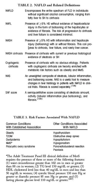
Definitions
For defining NAFLD, there must be (1) evidence of hepatic steatosis (HS), either by imaging or histology, and (2) lack of secondary causes of hepatic fat accumulation such as significant alcohol consumption, long-term use of a steatogenic medication, or monogenic hereditary disorders (Table 1). In the majority of patients, NAFLD is commonly associated with metabolic comorbidities such as obesity, diabetes mellitus, and dyslipidemia. NAFLD can be categorized histologically into nonalcoholic fatty liver (NAFL) or nonalcoholic steatohepatitis (NASH; Table 2). NAFL is defined as the presence of ≥5% HS without evidence of hepatocellular injury in the form of hepatocyte ballooning. NASH is defined as the presence of ≥5% HS and inflammation with hepatocyte injury (e.g., ballooning), with or without any fibrosis. For defining "advanced" fibrosis, this guidance document will be referring specifically to stages 3 or 4, that is, bridging fibrosis or cirrhosis.
SOME SELECTED HIGHLIGHTS - full text pdf attached
"Lifestyle modification consisting of diet, exercise, and weight loss has been advocated to treat patients with NAFLD........Oxidative stress is considered a key mechanism of hepatocellular injury and disease progression in subjects with NASH......Obesity (excessive body mass index [BMI] and visceral obesity) is the most common and well-documented risk factor for NAFLD. In fact, the entire spectrum of obesity, ranging from overweight to obese and severely obese, is associated with NAFLD.....Type 2 diabetes mellitus (T2DM): There is a very high prevalence of NAFLD in individuals with T2DM.....Dyslipidemia: High serum triglyceride (TG) levels and low serum high-density lipoprotein (HDL) levels are also common in patients with NAFLD. The prevalence of NAFLD in individuals with dyslipidemia attending lipid clinics has been estimated to be 50%.....initial reports suggested that compared to non-Hispanic whites, Hispanic individuals have a significantly higher prevalence of NAFLD, whereas non-Hispanic blacks have a significantly lower prevalence of NAFLD.[39]....One of the important surrogates for advanced liver disease is documentation of progressive hepatic fibrosis (HF)......long-term outcome of liver disease is the development of HCC. The current HCC incidence rate among NAFLD patients was determined to be 0.44 (range, 0.29-0.66) per 1,000 person-years.[16] In another study of patients with HCC, 54.9% of the HCC cases were related to HCV, 16.4% to alcoholic liver disease, 14.1% were related to NAFLD, and 9.5% to hepatitis B virus. However, it is estimated that the risk for developing HCC in NAFLD patients without cirrhosis is very small given the extremely large number of patients with NAFLD without cirrhosis within the general population.[61]......Vitamin E is an antioxidant and has been investigated as a treatment for NASH....There are also lingering concerns about the long-term safety of vitamin E....One meta-analysis suggested that doses of >800 IU/day were associated with increased all-cause mortality.....In a large RCT published in 2011, vitamin E administered at a dose of 400 IU/day was unexpectedly and unexplainably associated with a modest increase in the risk of prostate cancer (absolute increase of 1.6 per 1,000 person-years of vitamin E use),[189] and this risk may be modified by baseline selenium concentration[190] or genetic variants associated with vitamin metabolism.....Despite these limitations, it can be summarized that (1) the use of vitamin E is associated with a decrease in aminotransferases in subjects with NASH, (2) studies in which histological endpoints were evaluated indicate that vitamin E results in improvement in steatosis, inflammation, and ballooning and resolution of SH in a proportion of nondiabetic adults with NASH, and (3) vitamin E did not have an effect on HF. In the PIVENS clinical trial,[167] the pure form of rrr α-tocopherol was orally administered at a dose of 800 IU/day for 96 weeks. The primary endpoint was achieved in a significantly greater number of participants receiving vitamin E compared to placebo (42% vs. 19%; P < 0.001, number needed to treat = 4.4). In the Treatment of Nonalcoholic Fatty Liver Disease in Children trial (TONIC), which tested vitamin E (800 IU/day) or metformin (500 mg twice-daily) against placebo in children with biopsy-proven NAFLD, resolution of NASH was significantly greater in children treated with vitamin E than in children treated with placebo (58% vs. 28%; P = 0.006).[183] Two recent meta-analyses reported significant histological benefits with vitamin E in patients with NASH.[184, 185].......
---------------------------------
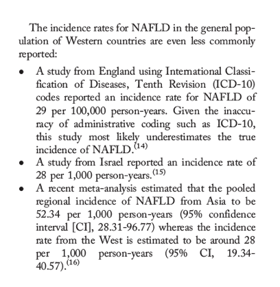
Prevalence of NAFLD in High-Risk Groups
Features of metabolic syndrome (MetS) are not only highly prevalent in patients with NAFLD, but components of MetS also increase the risk of developing NAFLD.[16, 18-20] This bidirectional association between NAFLD and components of MetS has been strongly established. In this context, Table 3 provides a list of the established conditions (obesity, type 2 diabetes, hypertension, and dyslipidemia) and emerging conditions (sleep apnea, colorectal cancer, osteoporosis, psoriasis, endocrinopathies, and polycystic ovary syndrome independent of obesity) that are associated with NAFLD.[21, 22]
⋅Obesity (excessive body mass index [BMI] and visceral obesity) is the most common and well-documented risk factor for NAFLD. In fact, the entire spectrum of obesity, ranging from overweight to obese and severely obese, is associated with NAFLD. In this context, the majority (>95%) of patients with severe obesity undergoing bariatric surgery will have NAFLD.[23, 24]
⋅Type 2 diabetes mellitus (T2DM): There is a very high prevalence of NAFLD in individuals with T2DM. In fact, some studies have suggested that around one third to two thirds of diabetic patients have NAFLD.[18, 25-27] It is also important to remember the importance of bidirectional association between NAFLD and T2DM. In this context, T2DM and NAFLD can develop almost simultaneously in a patient, which confounds the prevalence of NAFLD in patients with T2DM or the prevalence of T2DM in patients with NAFLD. Nevertheless, this association and its bidirectional causal relationship require additional investigation.[28]
⋅Dyslipidemia: High serum triglyceride (TG) levels and low serum high-density lipoprotein (HDL) levels are also common in patients with NAFLD. The prevalence of NAFLD in individuals with dyslipidemia attending lipid clinics has been estimated to be 50%.[29, 30] In a large, cross-sectional study conducted among 44,767 Taiwanese patients who attended a single clinic, the enrollees were stratified into four subgroups based on their total cholesterol to HDL-cholesterol and TG to HDL-cholesterol ratios. The overall prevalence rate of NAFLD was 53.76%; however, the NAFLD prevalence rate for those with the lowest total cholesterol to HDL-cholesterol and TG to HDL-cholesterol ratios was 33.41%, whereas the prevalence rate in the group with the highest ratios was 78.04%.
⋅Age, sex, and ethnicity: The prevalence of NAFLD may vary according to age, sex, and ethnicity.[31-39] In fact, both the prevalence of NAFLD and stage of liver disease appear to increase with age.[34-37]
----------------------------------------
Natural History and Outcomes of NAFLD
Over the past two decades, studies have reported the natural history of patients with NAFLD.[1, 19, 41-52] There is growing evidence that patients with histological NASH, especially those with some degree of fibrosis, are at higher risk for adverse outcomes such as cirrhosis and liver-related mortality.[1, 18, 19, 41-52] These studies have also shown the following:
⋅Patients with NAFLD have increased overall mortality compared to matched control populations without NAFLD.[53, 54]
⋅The most common cause of death in patients with NAFLD is cardiovascular disease (CVD), independent of other metabolic comorbidities.
⋅Although liver-related mortality is the 12th leading cause of death in the general population, it is the second or third cause of death among patients with NAFLD.[55]
⋅Cancer-related mortality is among the top three causes of death in subjects with NAFLD.[55]
⋅Patients with histological NASH have an increased liver-related mortality rate.[56, 57]
⋅In a recent meta-analysis, liver-specific and overall mortality rates among NAFLD and NASH were determined to be 0.77 per 1,000 (range, 0.33-1.77) and 11.77 per 1,000 person-years (range, 7.10-19.53) and 15.44 per 1,000 (range, 11.72-20.34) and 25.56 per 1,000 person-years (range, 6.29-103.80), respectively.[16]
⋅The incidence risk ratios for liver-specific and overall mortality for NAFLD were also determined to be 1.94 (range, 1.28-2.92) and 1.05 (range, 0.70-1.56), respectively.[16]
⋅The most important histological feature of NAFLD associated with long-term mortality is fibrosis; specifically, zone 3 sinusoidal fibrosis plus periportal fibrosis (stage 2) to advanced (bridging fibrosis [stage 3] or cirrhosis [stage 4]). These are independently predictive of liver-related mortality.[44, 58, 59]
⋅NAFLD is now considered the third-most common cause of hepatocellular carcinoma (HCC) in the United States, likely attributed to the enormous number of patients with the condition.[60] Given the growing epidemic of obesity, the incidence of NAFLD-related HCC has been shown to increase at a 9% annual rate.[61]
⋅Patients with NAFLD-related HCC are older, have a shorter survival time, more often have heart disease, and are more likely to die from their primary liver cancer than other HCC patients.[60]
⋅Around 13% of HCC reported from a study of patients from the Veteran Administration did not have cirrhosis. Among other factors, having NAFLD was independently associated with HCC in the absence of cirrhosis. This study confirms past small reports of HCC in NAFLD patients without cirrhosis.[62]
⋅It is important to recognize that most patients with cryptogenic cirrhosis may have what is considered "burned out" NAFLD.[63] This particular group of patients with cryptogenic cirrhosis have a disproportionately high prevalence of metabolic risk factors (T2DM, obesity, and MetS) that resemble patients with NAFLD, but the pathological assessment seldom reports histological features consistent with NASH or even steatosis in the presence of cirrhosis.[63, 64]
----------------------
Initial Evaluation of the Patient With Suspected NAFLD
The diagnosis of NAFLD requires that (1) there is HS by imaging or histology, (2) there is no significant alcohol consumption, (3) there are no competing etiologies for HS, and (4) there are no coexisting causes of CLD.
Common alternative causes of HS are significant alcohol consumption, hepatitis C, medications, parenteral nutrition, Wilson's disease (WD), and severe malnutrition (Table 1). When evaluating a patient with newly suspected NAFLD, it is important to exclude coexisting etiologies for CLD, including hemochromatosis, autoimmune liver disease, chronic viral hepatitis, alpha-1 antitrypsin deficiency, WD, and drug-induced liver injury.
Serological evaluation can uncover laboratory abnormalities in patients with NAFLD that do not always reflect the presence of another liver disease. Two examples of this are elevated serum ferritin and autoimmune antibodies. Mildly elevated serum ferritin is a common feature of NAFLD that does not necessarily indicate hepatic iron overload, though it can impact disease progression. Although the data are somewhat conflicting, serum ferritin >1.5 upper limit of normal (ULN) was associated with more advanced fibrosis in a retrospective cohort of 628 adults.[85] If serum ferritin and transferrin saturation are elevated in a patient with suspected NAFLD, genetic hemochromatosis should be excluded. Mutations in the HFE gene occur with variable frequency in patients with NAFLD, and the clinical significance is unclear.[86] Liver biopsy should be considered in the setting of high ferritin and a high iron saturation to determine the presence or extent of hepatic iron accumulation and to exclude significant hepatic injury in a patient with suspected NAFLD. Low titers of serum autoantibodies, particularly antismooth muscle and antinuclear antibodies, are common in patients with NAFLD and are generally considered to be an epiphenomenon of no clinical consequence, though they often require liver biopsy to exclude autoimmune disease. In a study of 864 well-characterized NAFLD subjects from the NASH Clinical Research Network (NASH CRN), significant elevations in serum autoantibodies (antinuclear antibodies >1:160 or antismooth muscle antibodies >1:40) were present in 21% and were not associated with more advanced disease or atypical histological features.[87]
While other diseases are being excluded, history should be carefully taken for the presence of commonly associated comorbidities, including central obesity, hypertension, dyslipidemia, diabetes or insulin resistance (IR), hypothyroidism, polycystic ovary syndrome, and obstructive sleep apnea......The diagnosis of NAFLD requires that (1) there is HS by imaging or histology, (2) there is no significant alcohol consumption, (3) there are no competing etiologies for HS, and (4) there are no coexisting causes of CLD.
Common alternative causes of HS are significant alcohol consumption, hepatitis C, medications, parenteral nutrition, Wilson's disease (WD), and severe malnutrition (Table 1). When evaluating a patient with newly suspected NAFLD, it is important to exclude coexisting etiologies for CLD, including hemochromatosis, autoimmune liver disease, chronic viral hepatitis, alpha-1 antitrypsin deficiency, WD, and drug-induced liver injury.
Guidance Statements:
7. When evaluating a patient with suspected NAFLD, it is essential to exclude competing etiologies for steatosis and coexisting common CLD.
8. In patients with suspected NAFLD, persistently high serum ferritin, and increased iron saturation, especially in the context of homozygote or heterozygote C282Y HFE mutation, a liver biopsy should be considered.
9. High serum titers of autoantibodies in association with other features suggestive of autoimmune liver disease (>5 ULN aminotransferases, high globulins, or high total protein to albumin ratio) should prompt a work-up for autoimmune liver disease.
10. Initial evaluation of patients with suspected NAFLD should carefully consider the presence of commonly associated comorbidities such as obesity, dyslipidemia, IR or diabetes, hypothyroidism, polycystic ovary syndrome, and sleep apnea.
--------------------------
Noninvasive Assessment of Steatohepatitis and Advanced Fibrosis in NAFLD
The natural history of NAFLD is fairly dichotomous�NAFL is generally benign, whereas NASH can progress to cirrhosis, liver failure, and liver cancer. Liver biopsy is currently the most reliable approach for identifying the presence of steatohepatitis (SH) and fibrosis in patients with NAFLD, but it is generally acknowledged that biopsy is limited by cost, sampling error, and procedure-related morbidity and mortality. Serum aminotransferase levels and imaging tests, such as ultrasound, computed tomography (CT), and MR, do not reliably reflect the spectrum of liver histology in patients with NAFLD. Therefore, there has been significant interest in developing clinical prediction rules and noninvasive biomarkers for identifying SH in patients with NAFLD, but their detailed discussion is beyond the scope of this practice guidance.[47]
NONINVASIVE QUANTIFICATION OF HEPATIC STEATOSIS (HS) IN NAFLD
Some studies suggest that degree of steatosis may predict the severity of histological features (e.g., ballooning and SH)[88] and the incidence and prevalence of diabetes in patients with NAFLD.[89-91] MR imaging, either by spectroscopy[92] or by proton density fat fraction,[93, 94] is an excellent noninvasive modality for quantifying HS and is being widely used in NAFLD clinical trials.[95] The use of TE to obtain continuous attenuation parameters is a promising tool for quantifying hepatic fat in an ambulatory setting.[74, 96] However, the utility of noninvasively quantifying HS in patients with NAFLD in routine clinical care is limited.
NONINVASIVE PREDICTION OF STEATOHEPATITIS (SH) IN PATIENTS WITH NAFLD
The presence of MetS is a strong predictor for the presence of SH in patients with NAFLD.[47, 97-100] Although NAFLD is highly associated with components of MetS, the presence of increasing an number of metabolic diseases, such as IR, type 2 diabetes, hypertension dyslipidemia, and visceral obesity, seems to increase the risk of progressive liver disease.[16, 18, 41] Therefore, patients with NAFLD and multiple risk factors such as T2DM and hypertension are at the highest risk for adverse outcomes.[20, 101] Circulating levels of cytokeratin-18 fragments have been investigated extensively as novel biomarkers for the presence of SH in patients with NAFLD.[47, 102, 103] This test is currently not available in a clinical care setting.
NONINVASIVE ASSESSMENT OF ADVANCED FIBROSIS IN PATIENTS WITH NAFLD
The commonly investigated noninvasive tools for the presence of advanced fibrosis in NAFLD include clinical decision aids (e.g., NAFLD fibrosis score, FIB-4 index, aspartate aminotransferase [AST] to platelet ratio index [APRI]), serum biomarkers (Enhanced Liver Fibrosis [ELF] panel, Fibrometer, FibroTest, and Hepascore), or imaging (eg, TE, MR elastography [MRE], acoustic radiation force impulse imaging, and supersonic shear wave elastography).[104]
The NFS is based on six readily available variables (age, BMI, hyperglycemia, platelet count, albumin, and AST/ALT ratio) and is calculated using the published formula (http://gihep.com/calculators/hepatology/nafld-fibrosis-score/). In a meta-analysis of 13 studies consisting of 3,064 patients,[47] the NFS had an area under the receiver operating curve (AUROC) of 0.85 for predicting advanced fibrosis (i.e., bridging fibrosis with nodularity or cirrhosis). A score <-1.455 had 90% sensitivity and 60% specificity to exclude advanced fibrosis, whereas a score >0.676 had 67% sensitivity and 97% specificity to identify the presence of advanced fibrosis. FIB-4 index (http://gihep.com/calculators/hepatology/fibrosis-4-score/) is an algorithm based on platelet count, age, AST, and ALT that offers dual cut-off values (patients with score <1.45 are unlikely, whereas patients with score >3.25 are likely to have advanced fibrosis).[104] A recent study that compared various risk scores and elastography (MR as well as TE) against liver histology showed that NFS and FIB-4 were (1) better than other indices such as BARD, APRI, and AST/ALT ratio and (2) as good as MRE for predicting advanced fibrosis in patients with biopsy-proven NAFLD.[105]
The ELF panel consists of plasma levels of 3 matrix turnover proteins (hyaluronic acid, tissue inhibitor of metalloproteinase 1, and N-terminal procollagen III-peptide) had an AUROC of 0.90 with 80% sensitivity and 90% specificity for detecting advanced fibrosis (bridging fibrosis or cirrhosis).[106] This panel has been recently approved for commercial use in Europe, but is not available for clinical use in the United States.
VCTE (FibroScan), which measures liver stiffness noninvasively, was recently approved by the U.S. Food and Drug Administration (FDA) for use in both adults and children with liver diseases. Two recent studies investigated the performance of VCTE in patients with suspected NAFLD using an M probe. Tapper et al. reported on the performance of VCTE in 164 patients with biopsy-proven NAFLD (median BMI, 32.2 kg/m2) from the United States.[107, 108] The optimal liver stiffness measurement cutoff for advanced fibrosis was 9.9 kilopascals with 95% sensitivity and 77% specificity. The AUROC for detecting advanced fibrosis was 0.93 (95% CI, 0.86-0.96). Interestingly, in 27% of the participants the VCTE yielded unreliable results.[107] Imajo et al. reported on the performance of VCTE using an with M probe in 142 Japanese patients with biopsy-proven NAFLD (mean BMI, 28.1 kg/m2).[108] The failure rate for VCTE in this cohort was 10.5%. The AUROC for VCTE for identifying advanced fibrosis (bridging fibrosis and cirrhosis) was 0.88 (95% CI, 0.79-0.97). The NASH CRN recently reported its experience with VCTE in 511 patients with biopsy-proven NAFLD (mean BMI, 33.6 kg/m2) across eight clinical centers in the United States, using a machine-guided protocol with either an M + or XL + probe.[109] Failure rate for obtaining a reliable liver stiffness measurement was 2.6%. MRE is excellent for identifying varying degrees of fibrosis in patients with NAFLD.[110, 111] In the study by Imajo et al., MRE performed better than VCTE for identifying fibrosis stage 2 or above, but they both performed equally well in identifying fibrosis stage 3 or above (i.e., bridging fibrosis). AUROCs for TE and MRE were 0.88 and 0.89, respectively.
Recent genome-wide association studies have associated several genetic polymorphisms, notably PNPLA-3 variants, with SH and advanced fibrosis in patients with NAFLD.[112-121] However, testing for these genetic variants in routine clinical care is currently not advocated.
Guidance Statements:
11. In patients with NAFLD, MetS predicts the presence of SH, and its presence can be used to target patients for a liver biopsy.
12. NFS or FIB-4 index are clinically useful tools for identifying NAFLD patients with higher likelihood of having bridging fibrosis (stage 3) or cirrhosis (stage 4).
13. VCTE or MRE are clinically useful tools for identifying advanced fibrosis in patients with NAFLD.
|
| |
|
|
|
|
|