 |
|
|
| |
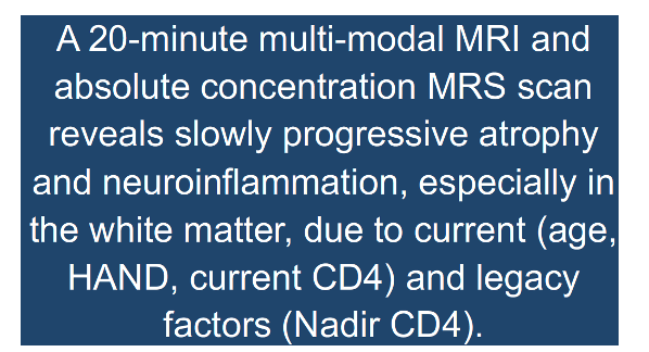
MRI With T2 Maps and Spectroscopy Show Chronic Progressive Brain Damage Despite HIV Suppression
|
| |
| |
CROI 2024 March 3-6 Denver
Bruce J. Brew1 3, David Jakabek 1, Lauriane Juge2 3, Kurt Lancaster1 3, Caroline D. Rae2 3, Lucette A Cysique1 3
1St. Vincent’s Hospital, Sydney, NSW, Australia, 2Insittution 2 Neuroscience Research Australia, Sydney, NSW, Australia, 3 The University of New South Wales, Sydney, Australia
program abstract
Background: We used a 20-minute absolute single-voxel proton magnetic resonance spectroscopy (MRS) scan with relaxometry (T1 and T2 maps) to better quantify water as a reference for major brain metabolites in virally suppressed people living with HIV (PWH) who are aging and were assessed for HIV immune markers and cognitive functions.
Methods: 39 PWH, all male (mean age: 53 ± 14, 60+ years old: 35%, HIV duration: 18 years, 20% nadir CD4 <200 cp/mL) were enrolled into a prospective study investigating brain metabolites during long-term viral suppression. They completed a baseline and a two-year follow-up clinical visit for HIV biomarkers (CD4 and CD8 count, nadir CD4, HIV duration), a neuropsychological assessment, a structural brain MRI scan, and a 20-minute single-voxel MRS in the frontal white matter, right caudate, and posterior cingulate with whole brain T1 and T2 mapping to derive the absolute concentration of major brain metabolites. MRS data were fitted with the spant R package and scaled using partial volumes (derived from Freesurfer and FSL MRS tools structural volumes). Mixed effect models assessed associations and longitudinal changes to T1, T2, and metabolite concentrations with age, baseline HIV biomarkers, and HIV-associated cognitive disorder (HAND).
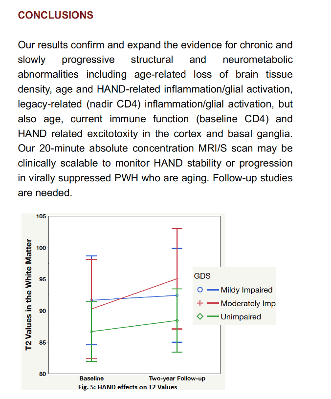
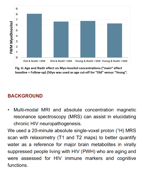
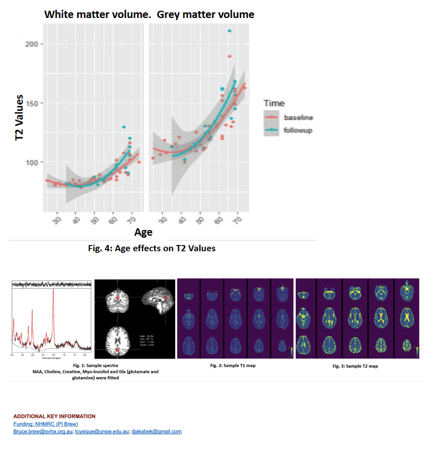
Results: Higher T2, but not T1 values, increased with age in white and grey matter (p < 0.002). HAND was associated with higher T2 values over time (p < 0.03). Older adults with HAND had higher T2 values than older adults without HAND (p < 0.04). Myo-Inositol concentration increased in all brain regions as a function of increased age (p < 0.01). There was a significant age-by-nadir CD4 interaction in frontal white matter for myo-Inositol (p < 0.001). Lower baseline CD4+ cell count and HAND were associated with lower glutamate in the cingulate (p = 0.04) and caudate (p = 0.003) of older participants, respectively.
Conclusion: Our results confirm and expand the evidence for chronic and slowly progressive structural and neurometabolic abnormalities including age-related loss of brain tissue density, age and HAND-related inflammation/glial activation, legacy-related (nadir CD4) inflammation/glial activation, but also age, current immune function (baseline CD4) and HAND related excitotoxicity in the cortex and basal ganglia. Our 20-minute absolute concentration MRI/S scan may be clinically scalable to monitor HAND stability or progression in virally suppressed PWH who are aging.
|
| |
|
|
|
|
|