 |
|
|
| |
Low Levels of HIV-1 in CSF During ART Are Associated
With Neurocognitive Impairment and Inflammation
|
| |
| |
CROI 2024 March 3-6 Denver
Laura P. Kincer1, Sarah B. Joseph1, Jessica R. Keys1, Natalie M. Bowman1, Chris Evans1, Alyssa Vecchio1, Serena Spudich2, Magnus Gisslén3, Prema Menezes1, Frank Maldarelli4, Robert Gorelick4, Joseph J. Eron1, Richard W. Price5, Ronald Swanstrom1
1University of North Carolina at Chapel Hill, Chapel Hill, NC, USA, 2Yale University, New Haven, CT, USA, 3Sahlgrenska Academy at the University of Gothenburg, Gothenburg, Sweden, 4National Institute of Health, Frederick, MD, USA, 5University of California San Francisco, San Francisco, CA, USA
Program Abstract:
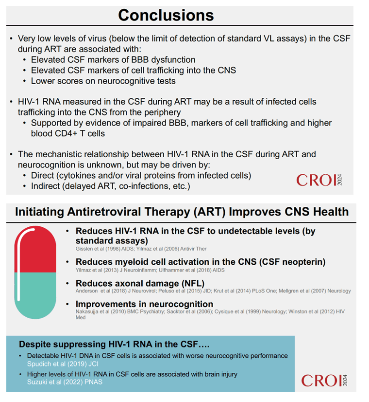
Background: Antiretroviral therapy (ART) typically reduces HIV-1 RNA to below the limit of detection of standard vial load (VL) assays and facilitates immune reconstitution, but neurocognitive impairment (NCI) persists in many people on ART. Previous studies have suggested that higher HIV-1 cell-associated DNA and cell-free RNA levels in cerebrospinal fluid (CSF) are positively associated with neurocognitive impairment (NCI), but the mechanisms driving this association are unknown.
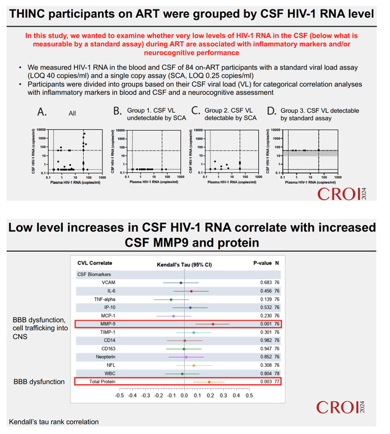
Methods: In a cross-sectional cohort of participants (N=78) who were on ART for at least a year and lacked overt neurologic symptoms, we examined whether the amount of HIV-1 RNA in cerebrospinal fluid (CSF) during ART is associated with elevated inflammation and/or NCI. We measured neurocognition by an 11-test battery and also measured plasma and CSF VLs (by both standard [Abbott Real Time assay, limit of detection (LOD) 40 cps/ml] and single copy assays [SCA, HHMCgag assay, LOD 0.25 cps/ml]), cell counts, and 12 inflammatory biomarkers.
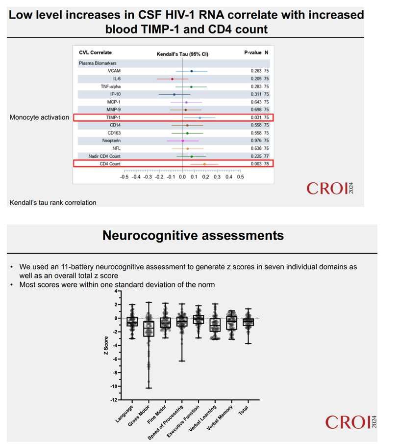
Results: ART regimens were NNRTI- (34%), PI- (33%), and InSTI-based (29%). Median blood CD4+ T cell counts, nadir blood CD4+ T cell counts, and CSF WBC counts were 506 cells/μl, 127 cells/μl, and 1 cell/μl, respectively. The cohort was divided into three groups with increasing HIV-1 RNA levels in CSF (median, IQR cp HIV-1 RNA/ml): (1) undetectable by all assays, (2) detectable by SCA only (0.33, 0.27-1.2 by SCA), (3) detectable by standard assay (40, 40-40 by standard assay). Using Kendall's tau rank correlation (Figure 1), we observed that levels of HIV-1 RNA in CSF were positively correlated with CSF MMP9
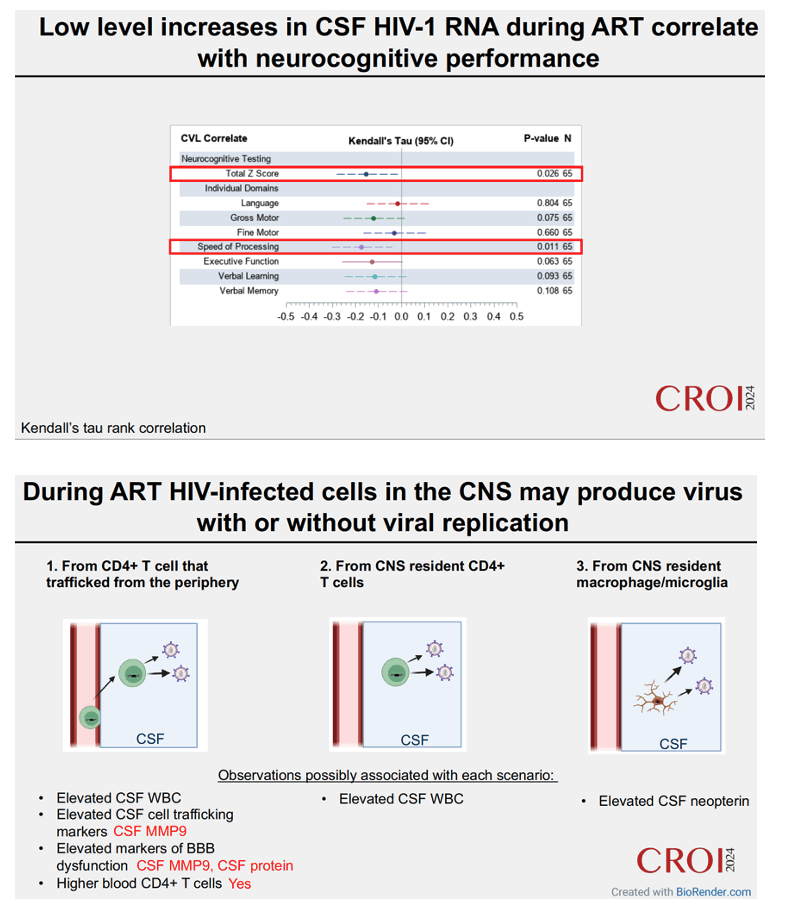
(p=0.001), CSF protein (p=0.001), plasma TIMP1 (p=0.031) and blood CD4+ T cell count (p=0.003) and negatively correlated with total neurocognition z score (p=0.026) and speed of processing z score (p=0.011).
Conclusion: We observed that during ART small increases in HIV-1 RNA that do not reach the level of treatment failure or CSF escape are associated with increased immune activation (CSF MMP9, plasma TIMP1), blood-brain barrier disruption (CSF protein) and neurocognitive impairment (total z and speed of processing z). This contributes to the growing evidence that persistent exposure to HIV-1 in the CNS during ART is associated with NCI and suggests that inflammation may play an important role in this process.
|
| |
|
|
|
|
|