 |
|
|
| |
SCH503034 & NM283 Not Cross-Resistant
|
| |
| |
Reported by Jules Levin
EASL, April 2007, Barcelona, Spain
"Combination Of Two Hepatitis C Virus Inhibitors, SCH 503034 (Boceprevir) And NM107 (Active Moiety of Valopicitabine), Provides Enhanced Anti-Replicon Activity And Suppresses Emergence Of Resistant Replicons"
R. Ralston1, V. Bichko2, R. Chase1, M. LaColla2, L. Lallos2, A. Skelton1, M. Soubasakos2, M. Tausek2, X. Tong1, D. Standring2
1Virology, Schering-Plough Research Institute, Kenilworth, NJ, USA; 2Virology, Idenix Pharmaceuticals, Cambridge, MA, USA
AUTHOR CONCLUSIONS
Combination treatment with SCH 503034 (boceprevir) plus NM107 (active moiety of valopicitabine) was additive in reducing levels of replicon protein and RNA levels.
Cross-resistance to either compound was not observed.
Reduction in emergence of resistant colonies was enhanced by the combination compared to either drug used alone.
No cytotoxicity was observed.
ABSTRACT
Background: Combinations of small-molecule antiviral agents for the treatment of hepatitis C, particularly those with different molecular targets, offer the potential for enhanced suppression of hepatitis C virus (HCV) replication
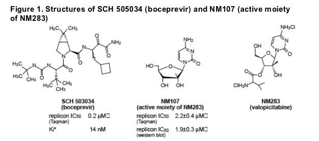
and reduced emergence of resistance. SCH 503034 (boceprevir) (Schering-Plough), a NS3 protease inhibitor, and NM283 (valopicitabine) (Idenix/Novartis), a nucleoside NS5B polymerase inhibitor, have demonstrated antiviral activity in clinical studies, although resistant viral variants have been detected. The combined antiviral effects of SCH 503034 (boceprevir) and NM107 (the active moiety of NM283 [valopicitabine]) were assessed using cell culture replicon studies.
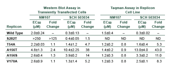
Methods: The combined antiviral effect of SCH 503034 (boceprevir) and NM107 was evaluated in genotype 1b HCV replicon cells by real time RT-PCR or ELISA. Possible cross-resistance of these compounds was evaluated using replicon variants carrying single amino acid substitutions (T54A, A156S, V170A, A156T) in the HCV protease that reduce susceptibility to SCH 503034 (boceprevir), and NM107-resistant replicons carrying the S282T substitution in the HCV polymerase. These substitutions in the HCV protease and polymerase reduced inhibitory potency 5- to 125-fold for SCH 503034 (boceprevir) and NM107 respectively.
Results: The combination of SCH 503034 (boceprevir) and NM107 showed dose-dependent enhancement of replicon inhibition, compared with the effect of each inhibitor used alone. No cytotoxicity was observed. In cross-resistance
studies, NM107 showed similar antiviral activity (EC50 1.5-2 mM) against wild-type and SCH 503034 (boceprevir)-resistant replicons. SCH 503034 (boceprevir) showed similar activity (EC50 0.3-0.4 mM) against wildtype and NM107-resistant replicons. The combination of SCH 503034 (boceprevir) and NM107 significantly reduced the frequency of resistant colonies in a dose-dependent manner, compared to each inhibitor used alone.
Conclusions: The combination of SCH 503034 (boceprevir) and NM107 showed enhanced anti-replicon activity with no cross-resistance, suggesting that the clinical antiviral effects of this combination will be greater than the effects seen with monotherapy, with a greater genetic barrier to the selection of clinically resistant viral variants. These results strongly support clinical evaluation of this combination in patients with chronic hepatitis C.
RESULTS
Combination Treatment With SCH 503034 (boceprevir) and NM107:
Results of 3-day Assays in Genotype 1b Replicon Cell Lines
- Reductions in levels of both replicon RNA and protein were observed
- Effect of combination treatment was at least additive in short term assay
- Cross-resistance was not observed
- No cytotoxicity was observed
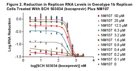
SCH 503034 (boceprevir) was added to replicon cells (Cl 16, genotype 1b) in a 10 point 1:2 serial dilution. NM107 was titrated into each concentration of SCH 503034 (boceprevir). The final concentrations of DMSO and fetal bovine serum are 1% and 10%, respectively. After three day treatment, the replicon RNA level is measured using real time PCR (Taqman assay). GAPDH RNA is used
as an endogenous control. Replicon RNA reduction is calculated as below:
- dCT = 5BCT-gapdhCT
- ddCT = dCT-dCTof no cpd control
- log RNA reduction = log (1/2ddCT)
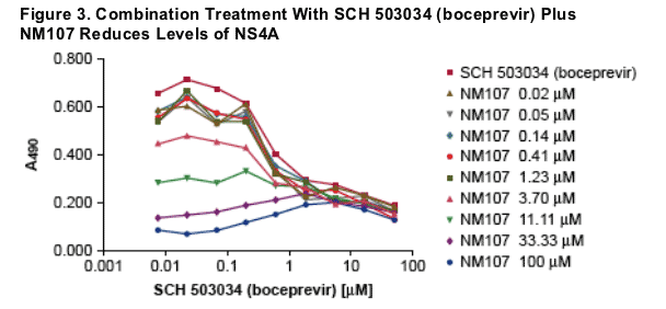
A stable Huh7 cell line harboring subgenomic HCV replicon (genotype 1b, Con1 isolate) was used. HCV replication was measured by NS4A ELISA. Data averaged from 5 independent experiments are shown.
Figure 4. Combination of NM107 & SCH 503034 (boceprevir) Have Additive Effects in Reducing NS4A Protein
Mathematical analyses of drug-drug interactions between NM107 and SCH 503034 (boceprevir) in the HCV replicon model, the Bliss independence model (A. MacSynergy II, 99.9% confidence level, B. CombiTool) and Loewe additivity model (C. CombiTool) were used in this study. For B and C, dotted lines represent 25% deviation from the calculated additive effect (midline).
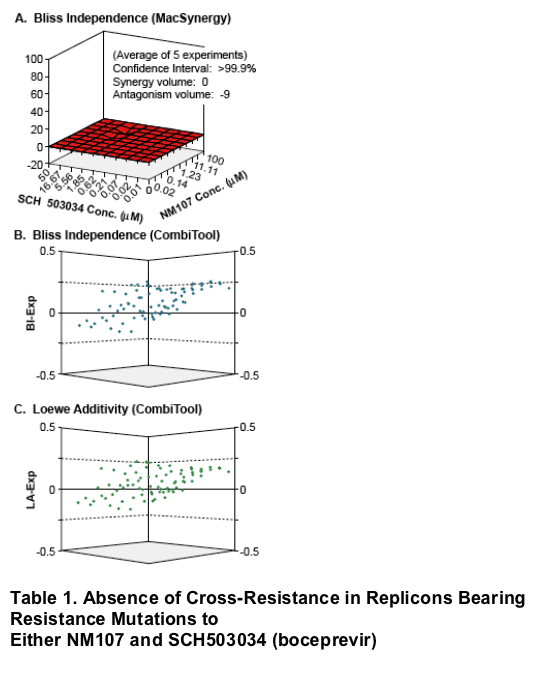
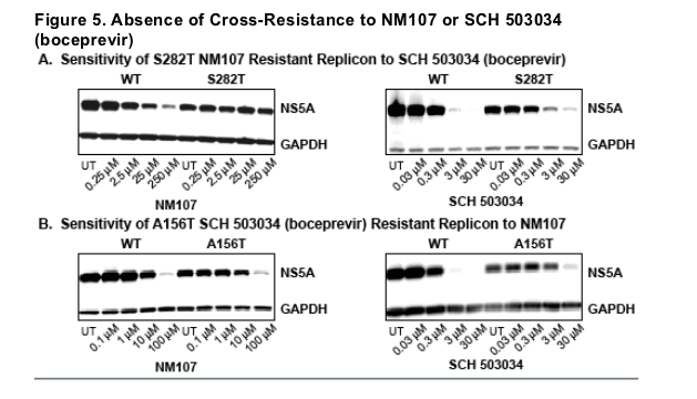
Cross-resistance of HCV replicons was assayed by two methods. For the western blot analysis, HCV replicon RNA was transiently transfected into Huh7 cells, followed by NS5A quantification (see Figure 5, below). For the Taqman assay, Huh7 cells stably transfected with replicons bearing the indicated resistance mutations to SCH 503034 (boceprevir). Data averaged from two experiments are shown.
A representative picture of the Western blot analyses. NS5A protein was quantified to measure HCV replication. A cellular protein, GAPDH, was used for normalization.
Combination treatment with SCH 503034 (boceprevir) and NM107: results of 2-week assays in genotype 1 replicon cell lines
- Combination treatment reduces the emergence of resistant colonies compared
to either drug used alone All cell cultures were split once during a 27-day drug treatment, followed by crystal violet staining. Results of a representative
experiment are shown.
All cell cultures were split once during a 27-day drug treatment, followed by crystal violet staining. Results of a representative experiment are shown.
|
| |
|
|
|
|
|