 |
|
|
| |
Lipid steatosis, metabolic variables and body composition in the setting of HIV/HCV coinfection; findings from a large randomised trial (APRICOT)
|
| |
| |
Reported by Jules Levin
EASL, April 2007, Barcelona, Spain
M. Rodriguez-Torres,1 S. Govindarajan,2 R. Sola,3 N. Clumeck,4 E. Lissen,5 M. Pessoa,6 P. Buggisch,7 J. Main,8 J. DePamphilis,9 D.T. Dieterich10
1Fundacion de Investigacion de Diego, Santurce, Ponce School of Medicine Puerto Rico; 2Rancho Los Amigos Medical Center, Downey, CA, USA; 3Universitat Autonoma de Barcelona, Barcelona, Spain;
4CHU Saint-Pierre, Brussels, Belgium; 5Virgen del Rocio University Hospital, Seville, Spain; 6Instituto de Infectologia Emilio Ribas, Sao Paulo, Brazil; 7Universitatsklinik Eppendorf, Hamburg, Germany;
8St Mary's Hospital, London, UK; 9Roche, Nutley, NJ, USA; 10Mount Sinai School of Medicine, New York, NY, USA
INTRODUCTION
Hepatic steatosis is common in patients with chronic hepatitis C virus (HCV) infection and is associated with accelerated fibrosis progression. This phenomenon has not been well studied in patients with HIV-HCV co-infection.
Baseline and end-of-follow-up liver biopsies were collected in a subset of patients enrolled in the AIDS PEGASYS Ribavirin International Co-infection Trial (APRICOT).[1] Treatment for 48 weeks with the combination of peginterferon alfa-2a (40KD) (PEGASYS) plus ribavirin (COPEGUS) produced an overall sustained virological response (SVR) rate of 40% in this study. Thus, data from this trial provide an opportunity to study the phenomenon of hepatic steatosis, including the impact of viral eradication.
AUTHOR CONCLUSIONS
This analysis adds to the evidence that body weight, body mass index (BMI) and non-specific lipodeposition are important prognostic factors associated with steatosis. (note from Jules: glucose, HDL cholesterol, ALT, and hip circumference, triglycerides, BMI, and nukes [AZT, ddI, d4T, ddC] all associated here as predictive factors for steatosis, see tables below).
The significant association between steatosis and HCV genotype 3 infection and with higher HCV RNA levels, and the resolution of steatosis in genotype 3 patients who achieve an SVR, suggests that HCV is critical to the pathogenesis of hepatic steatosis.
In contrast, the lack of resolution of steatosis in patients infected with non-genotype 3 patients who achieved an SVR suggests that there is the potential for further liver damage in these individuals.
Importantly, there was no association seen between steatosis and the achievement of SVR.
Further study is needed to explore these complex relationships.
METHODS
The study design, primary results and histological outcomes of APRICOT have been published elsewhere.[1,2]
Patients
Treatment-naive adults with HIV-HCV co-infection, quantifiable serum HCV RNA levels (>600 IU/mL), elevated serum analine aminotransferase (ALT) levels, a liver biopsy obtained within the previous 15 months and compensated liver disease were eligible for APRICOT.[1]
Patients were required to have stable HIV disease status and must have either been receiving stable antiretroviral therapy (ART) for 36 weeks prior to enrolment or had not been receiving ART for 38 weeks prior to randomisation with no plans to start within the next 6 weeks.
Study design
Patients were randomised to 48 weeks of treatment with peginterferon alfa-2a (40KD) 180 _g/week given alone or in combination with ribavirin 800 mg/day, or conventional interferon alfa-2b 3 MIU tiw plus ribavirin 800 mg/day.
SVR, the primary efficacy end point, was defined as undetectable serum HCV RNA (<50 IU/mL) at the end of a 24 week untreated follow-up period (i.e., study week 72).
The subset of patients considered in this analysis includes those that consented to an end-of-follow-up liver biopsy (all centres were invited to participate). Paired pretreatment and end-of-follow-up biopsy specimens were submitted to a central study pathologist for reading. Steatosis was defined as >5% of hepatocytes with fatty changes per low power field.
Anthropomorphic measurements of the neck, arm waist, hip, thigh and chest were obtained in a subset of patients.
According to American Heart Association criteria, the metabolic syndrome was defined as the presence of 33 or more of: waist circumference >102 cm in men, or >88 cm in women; serum triglycerides .150 mg/dL; HDL-cholesterol
<40 mg/dL in men, or <50 mg/dL in women; diastolic/systolic blood pressure >130/85 mm Hg; fasting plasma glucose >100 mg/dL.
RESULTS
Paired biopsy specimens from 33% (283/860) of patients were submitted and analysed for steatosis.
The prevalence of steatosis among patients with paired biopsies was 23% (65/283).
The prevalence of metabolic syndrome was 24% (67/277).
A higher proportion of patients with steatosis had concurrent metabolic syndrome at baseline (25/65, 38%) than patients without steatosis (42/212, 20%). (note from Jules: in other words the worse one's metabolics the more likely to have steatosis, so steatosis is not necessarily associated with ART but the degree of metabolic abnormality).
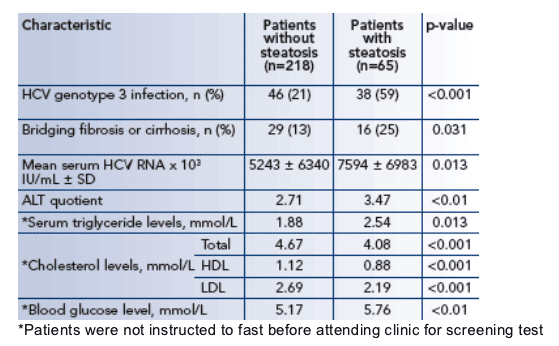
Significant differences in baseline characteristics between patients with and without steatosis are presented in Table 1.
Table 1. Baseline characteristics of patients with and without
steatosis (n=283)
Patients with steatosis had significantly more often genotype 3, more bridging fibrosis or cirrhosis, higher HCV RNA, higher ALT quotient, higher triglycerides, lower total cholesterol, LDL and abnormal HDL, and higher glucose (fasting not required).
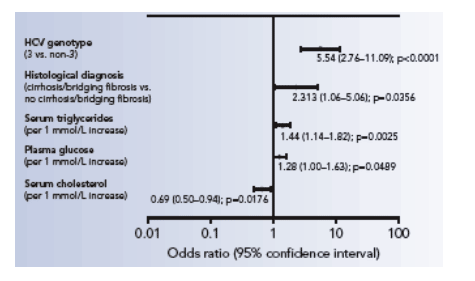
Several multivariate regression analyses were developed, the first of which included all patients with available data (Figure 1).
Factors independently associated with steatosis
All patients at baseline (n=282)
Serum cholesterol per 1 mmol/L increase: 0.69, p=0.0176; glucose: per 1 mmol/L increase: 1.28, p=0.0489; triglycerides per 1 mmol/L increase: 1.44, p=0.0025; histological diagnosis (cirrhosis/bridging fibrosis vs no cirrhosis/bridging fibrosis): 2.313, p=0.0356; HCV genotype (3 vs non-3): 5.54, p<0.0001.
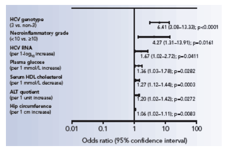
Factors independently associated with steatosis
Includes patients with HDL and anthropomorphic data at baseline (n=258)
Hip circumference per 1 cm increase: 1.06, p=0.0083; ALT quotient per 1 unit increase: 1.20, p=0.0272; HDL cholesterol per 1 mmol/L decrease: 1.27, p=0.0003; glucose per 1 mmol/L increase: 1.36, p=0282; HCV RNA per 1 log increase: 1.67, p=0.0411; necroinflammatory grade <10 vs >10: 4.27, p=0.0161; HCV genotype 3 vs non-3: 6.41, p<0.0001.
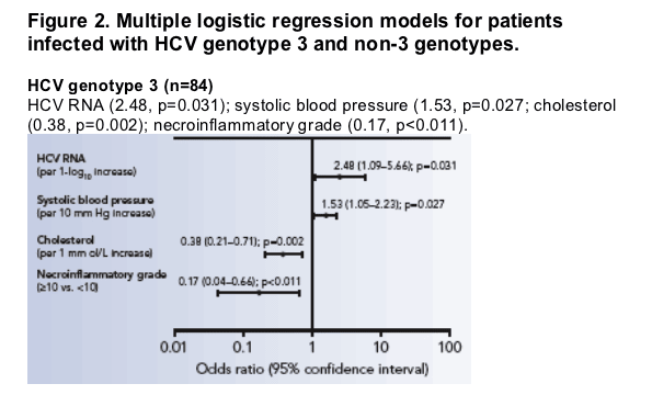
HCV genotype (3 vs. non-3) was the strongest predictor of steatosis, so separate logistic regression models were developed for patients with genotype 3 and those with non-3 genotypes (Figure 2). There were marked differences in the factors that were retained in the final models.
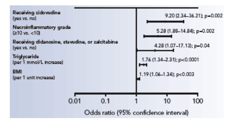
Figure 2. Multiple logistic regression models for patients
infected with HCV genotype 3 and non-3 genotypes.
HCV genotype 3 (n=84)
HCV RNA (2.48, p=0.031); systolic blood pressure (1.53, p=0.027; cholesterol (0.38, p=0.002); necroinflammatory grade (0.17, p<0.011).
HCV non-genotype 3 (n=198)
Receiving AZT (9.20, p=0.002); necroinflammatory grade (5.28, p=0.002); receiving ddI, d4T or ddC (4.28, p=0.04); triglycerides per 1 mmol/L increase (1.76, p<0.0001); BMI per 1 unit increase (1.19, p<0.003).
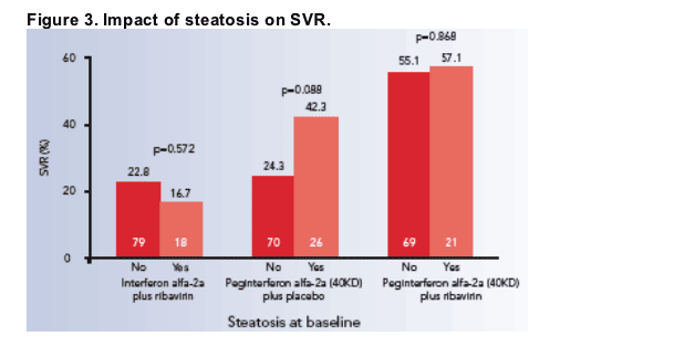
The presence of steatosis at baseline did not significantly affect the probability of achieving an SVR in any of the three treatment groups (Figure 3). For this reason, data from the three treatment groups were combined for the purposes of examining the influence of an SVR on steatosis within particular genotypes.
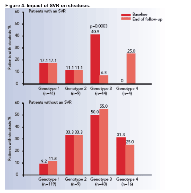
Eradication of HCV genotype 3 infection (i.e. SVR) was associated with a significant reduction of steatosis (Figure 4). This was not the case in patients infected with other genotypes. In non-responders there was no change in the prevalence of steatosis between baseline and end of follow-up.
REFERENCES
1. Torriani FJ, Rodriguez-Torres M, Rockstroh JK, et al. Peginterferon alfa-2a plus ribavirin for chronic hepatitis C virus infection in HIV-infected patients. N Engl J Med 2004; 351: 438-50.
2. Lissen E, Clumeck N, Sola R, et al. Histological response to pegIFNalpha-2a (40KD) plus ribavirin in HIV-hepatitis C virus co-infection. AIDS 2006; 20: 2175-81.
|
| |
|
|
|
|
|