 |
|
|
| |
Gut-associated Lymphoid Tissue Fibrosis Is Associated with CD4+ T Cell Activation and Poor HIV-specific CD8+ T cell Responses During Suppressive Antiretroviral Therapy
|
| |
| |
Reported by Jules Levin
CROI 2011 Boston March 2
Peter W. Hunt1, Hiroyu Hatano1, Ma Somsouk1, Elizabeth Sinclair1, Lorrie Epling1, Lee Gilman1, Jeffrey N. Martin1, Steven G. Deeks1, and Timothy Schacker2 1University of California, San Francisco; 2University of Minnesota, Minneapolis
Background
The HIV-specific CD8+ T cell response may be important in eliminating HIV-infected cells in future eradication strategies.
However, many HIV+ individuals fail to maintain detectable HIV-specific CD8+ T cell responses despite continued release of HIV RNA from latently infected cells during suppressive antiretroviral therapy (ART).
HIV-associated lymphoid tissue fibrosis damages anatomic structures required for efficient antigen presentation and T cell survival.
We hypothesized that lymphoid tissue fibrosis might also limit the ability to maintain HIV-specific CD8+ T cells.
METHODS
Sampled HIV-infected participants maintaining plasma HIV RNA levels <75 copies/ml on antiretroviral therapy (ART) from the SCOPE cohort.
Assessed % activated (CD38+HLA-DR+), PD1+, HIV Gag-specific IFN-γ+ T cells in cryopreserved PBMC as part of a larger study on immunologic correlates of poor ART-mediated CD4+ T cell recovery (see poster #504).
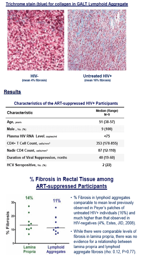
A subset of 9 of these individuals had flexible sigmoidoscopies performed with rectal mucosal biopsies. 4 biopsy pieces (each ~3mm) for each participant were fomalin fixed and paraffin embedded for histology.
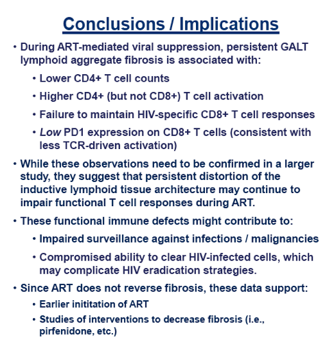
% Fibrosis: Rectal tissue was stained with a trichrome stain, which stains collagen a light blue color. The % area in lymphoid aggregates and in lamina propria occupied by collagen was assessed with quantitative image analysis (see representative figure below from Estes et al, JID, 2008):
|
| |
|
|
|
|
|