| |
Body composition changes after switching from protease inhibitors to raltegravir: SPIRAL-LIP substudy
|
| |
| |
Download the PDF here
Curran, Adriana; Martinez, Estebanb; Saumoy, Mariac; del Rio, Luisd; Crespo, Manuela; Larrousse, Mariab; Podzamczer, Danielc; Burgos, Joaquina; Lonca, Montseb; Domingo, Peree; Gatell, Jose Mariab; Ribera, Estebana
AIDS:
20 February 2012
aHospital Universitari Vall d'Hebron, Universitat Autonoma de Barcelona, Infectious Diseases Department
bHospital Clinic i Provincial, Infectious Diseases Department
cHospital Universitari de Bellvitge, HIV Unit, Infectious Diseases Department, L'Hospitalet de Llobregat
dCETIR, Nuclear Medicine Department
eHospital Universitari Santa Creu i Sant Pau, Universitat Autonoma de Barcelona, Infectious Diseases Department, Barcelona, Spain.
Abstract
Objective: To compare 48-week changes in body fat distribution and bone mineral density (BMD) between patients switching from a ritonavir-boosted protease inhibitor (PI/r) to raltegravir (RAL) and patients continuing with PI/r.
Design: Substudy of the prospective, randomized, open-label, multicenter SPIRAL study.
Methods: Patients were randomized (1 : 1) to continue with the PI/r-based regimen or switch to RAL, maintaining the rest of the treatment unchanged. Dual-energy X-ray absorptiometry and computed tomography scans were performed at baseline and after 48 weeks to measure body fat and bone composition, analyzing intragroup and intergroup differences.
Results: Eighty-six patients were included and 74 patients (39 RAL, 35 PI/r) completed the substudy. Significant increases in median [interquartile range (IQR)] visceral adipose tissue (VAT) [20.7 (-2.4 to 45.6) cm2, P = 0.002] and total adipose tissue (TAT) [21.4 (-1.3 to 55.4) cm2, P = 0.013] were seen within the PI/r group. No significant changes in body fat were seen with RAL or between treatment groups. Regarding bone composition, total BMD [0.01 (0 to 0.02) g/cm2, P = 0.002], total hip BMD [0.01 (0 to 0.03) g/cm2, P = 0.015] and total hip T score [0.12 (-0.05 to 0.21) SD, P = 0.004] significantly increased with RAL, with no significant changes within the PI/r group. Differences between treatment groups were significant in femoral neck BMD [0.01 (-0.02 to 0.02) g/cm2, P = 0.032] and T score [0.01 (-0.18 to 0.18) SD, P = 0.016].
Conclusion: Although there were no significant changes in body fat between groups, maintaining a PI/r-based regimen was associated with a significant increase in VAT and TAT. Switching to RAL led to a significant increase in femoral neck BMD when comparing between groups.
----------------------------------
Body fat distribution
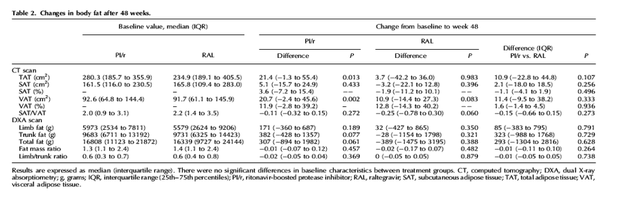
Body fat changes after 48 weeks are described in Table 2. No significant differences were seen within or between groups in weight or BMI (data not shown).
In the CT scans, a significant increase within the PI/r group was seen in median (IQR) VAT [20.7 (-2.4 to 45.6) cm2, P = 0.002] and TAT [21.4 (-1.3 to 55.4) cm2, P = 0.013], whereas no significant differences were seen within the RAL group [VAT 10.9 (-14.4 to 27.3) cm2, P = 0.083; TAT 3.4 (-42.2 to 36.0) cm2, P = 0.983] or between treatment arms (P = 0.333 for VAT and P = 0.107 for TAT). No significant differences were seen in SAT or SAT/VAT ratios within or between treatment groups (Table 2).
Regarding the DXA scans, there were small increases in limb fat with both regimens, whereas in trunk and total fat there was a nearly significant increase with PI/r (trunk fat 382 g, P = 0.077; total fat 307 g, P = 0.061) and a nonsignificant decrease with RAL (Table 2). None of the changes reached significance within or between groups.
There were no significant differences in body fat composition depending on the NRTI backbone used in the current treatment (data not shown).
Bone mineral density
There were no significant changes in BMD or T scores in any location with PI/r after 48 weeks. On the contrary, total BMD [0.01 (0 to 0.02) g/cm2, P = 0.002] and total hip BMD [0.01 (0 to 0.03) g/cm2, P = 0.015] increased significantly within the RAL group, as did the total hip T score [0.12 (-0.05 to 0.21) SD, P = 0.004]. T scores in femoral neck and spine (L1-L4) had a nearly significant increase with RAL (Table 3). When we compared both treatment arms, there were significant differences favoring RAL in femoral neck BMD and T scores (Table 3), without differences in the other locations.
There were no significant differences in BMD or T scores in either group depending on TDF use (data not shown).
There was no correlation between changes in body fat (by CT or DXA) and changes in BMD or T scores in any location (data not shown).
----------------------------
Introduction
Antiretroviral therapy (ART) has changed the evolution of HIV infection, decreasing overall and, particularly, AIDS-related morbidity and mortality. However, it has some drawbacks. One of the most feared adverse effects of ART is lipodystrophy, a body fat distribution alteration that includes lipoatrophy (fat loss in face, limbs and buttocks) and lipohypertrophy (fat accumulation in breasts, visceral and dorsocervical regions). It has been postulated that lipoatrophy and lipohypertrophy are two separate conditions with different risk factors [1,2]. Lipoatrophy has been related mainly to the duration of exposure to thymidine analogs (zidovudine and estavudine) [3-5], whereas lipohypertrophy pathogenesis is not so clearly established, although at least some protease inhibitors have been involved in its apparition [6]. Some studies have shown differences in visceral adipose tissue (VAT) in naive patients after starting different ritonavir-boosted protease inhibitor (PI/r)-based ART [7] and also after switching from one PI/r to another [8]. Other studies have compared body fat changes with PI/r versus non-nucleoside reverse transcriptase inhibitors (NNRTIs), with discordant results [9-11].
Raltegravir (RAL) is the first integrase inhibitor clinically available in the ART armamentarium. RAL has shown a better lipid profile than efavirenz in naïve patients in the STARTMRK trial [12], and switching from PI/r to RAL also has led to decreases in plasma lipids [13,14]. Regarding body fat distribution, the only available information is from the aforementioned STARTMRK study, in which RAL and efavirenz showed comparable fat gain at 96 weeks, both in trunk and arms [12]. There is no body composition data for RAL in patients with suppressed HIV infection or published studies comparing RAL with PI/r.
Decreased bone mineral density (BMD) is another growing concern for HIV-infected patients. The incidence of osteopenia/osteoporosis and bone fractures is higher in these patients compared to the general population [15,16]. Although HIV patients frequently have more classical risk factors for low BMD (e.g. low body mass index, smoking, sedentary lifestyle), both HIV infection and ART may also play a potential role. Tenofovir (TDF) has been associated with increased BMD loss in some studies [11,17] and there are conflicting data about decreased BMD and PI/r exposure [18-20]. There are no published data, to our knowledge, for RAL and BMD.
The aim of this study was to evaluate 48-week changes in body fat distribution and bone composition after switching from a PI/r to RAL compared to maintaining the PI/r.
Methods
Design
The SPIRAL trial (clinicaltrials.gov NCT00528892) is a prospective, randomized, open-label, multicenter study in which patients with suppressed HIV-1 infection on a PI/r regimen were randomized (1 : 1) to continue with the same treatment or change the PI/r to RAL 400 mg b.i.d., without modifying the rest of the ART. The details of the global study are described elsewhere [14].
The SPIRAL-LIP substudy was planned prior to the initiation of the SPIRAL study. Patients were enrolled consecutively from three sites in Barcelona, Spain, between June and December 2008. The primary endpoint was absolute change in VAT area. Secondary endpoints were changes in limb fat, trunk fat, total fat, total adipose tissue (TAT) area, subcutaneous adipose tissue (SAT) area and SAT/VAT ratios in body fat composition and changes in BMD and T scores in total body, spine (L1-L4) and hip (femoral neck and total hip) in bone composition.
The protocol was approved by the Ethics Committee at each participating site and a specific written informed consent for the substudy was obtained from all patients.
Procedures
At baseline and at week 48 whole body, lumbar and hip dual-energy X-ray absorptiometry (DXA) scans (Lunar DPXL, Madison, Wisconsin, USA) were performed to assess body fat and bone composition. At the same time points, a computed tomography (CT) scan of the abdomen (single cut at L4, 5 mm thick) was performed to assess VAT, SAT and TAT areas. Tissue areas (cm2) were calculated by summing specific tissue pixels and then multiplying by individual pixel surface area. The DXA and CT scans were performed in a single center following a standardized protocol and read by a single radiologist unaware of the patient's ART. The results of the DXA and CT scans were given to the physicians in care of the patients after the study was ended, so no specific actions were immediately derived from the results of the scans. No specific dietary supplementation was given to the patients.
Statistical analysis
A sample size of 74 evaluable patients was considered necessary to detect a difference of 7.6 cm2 in VAT (according to findings from ACTG 5224 study [11]) with an 80% power and a significance level-∝ of 0.05 using a two-sided test.
For quantitative variables, medians and interquartile ranges (IQRs) (25th-75th percentiles) were used as measures of central tendency and dispersion. The number of patients in each category and the corresponding percentages were given for qualitative variables. The changes from baseline were compared with the Wilcoxon's signed-rank and the McNemar tests for quantitative variables and the chi-squared test for qualitative variables, with the continuity correction for the chi-square when a subgroup included five or fewer patients. Comparisons between quantitative nonpaired variables were performed with the Mann-Whitney U test. Correlations were analyzed by Spearman's rank test. All statistical tests were two-tailed and were performed at a level of statistical significance of 0.05. SPSS 15.0 software for Windows (SPSS Inc., Chicago, Illinois, USA) was used for statistical analyses.
Results
Population
Eighty-six patients were included in the study. Twelve patients (7 in the PI/r arm and 5 in the RAL arm) did not complete the substudy: 1 was lost to follow-up and 11 patients did not have the DXA and CT scans performed at 48 weeks. There were no significant differences in baseline characteristics between patients who did and did not complete 48 weeks of follow-up and losses were balanced between treatment arms. Finally, 74 patients with 48 weeks data were analyzed. Of them, 39 had been randomized to switch to RAL and 35 to continue with the PI/r. Patient's baseline characteristics are described in Table 1. There were no significant differences in baseline characteristics between groups.
Body fat distribution
Body fat changes after 48 weeks are described in Table 2. No significant differences were seen within or between groups in weight or BMI (data not shown).
In the CT scans, a significant increase within the PI/r group was seen in median (IQR) VAT [20.7 (-2.4 to 45.6) cm2, P = 0.002] and TAT [21.4 (-1.3 to 55.4) cm2, P = 0.013], whereas no significant differences were seen within the RAL group [VAT 10.9 (-14.4 to 27.3) cm2, P = 0.083; TAT 3.4 (-42.2 to 36.0) cm2, P = 0.983] or between treatment arms (P = 0.333 for VAT and P = 0.107 for TAT). No significant differences were seen in SAT or SAT/VAT ratios within or between treatment groups (Table 2).
Regarding the DXA scans, there were small increases in limb fat with both regimens, whereas in trunk and total fat there was a nearly significant increase with PI/r (trunk fat 382 g, P = 0.077; total fat 307 g, P = 0.061) and a nonsignificant decrease with RAL (Table 2). None of the changes reached significance within or between groups.
There were no significant differences in body fat composition depending on the NRTI backbone used in the current treatment (data not shown).
Bone mineral density
There were no significant changes in BMD or T scores in any location with PI/r after 48 weeks. On the contrary, total BMD [0.01 (0 to 0.02) g/cm2, P = 0.002] and total hip BMD [0.01 (0 to 0.03) g/cm2, P = 0.015] increased significantly within the RAL group, as did the total hip T score [0.12 (-0.05 to 0.21) SD, P = 0.004]. T scores in femoral neck and spine (L1-L4) had a nearly significant increase with RAL (Table 3). When we compared both treatment arms, there were significant differences favoring RAL in femoral neck BMD and T scores (Table 3), without differences in the other locations.
There were no significant differences in BMD or T scores in either group depending on TDF use (data not shown).
There was no correlation between changes in body fat (by CT or DXA) and changes in BMD or T scores in any location (data not shown).
Discussion
With current ART, sustained virological control is a feasible therapeutic goal, and physicians treating HIV-infected patients aim for treatments with small long-term toxicity. Nowadays, two of the main concerns regarding side effects, especially in our growing old population, are body fat (with its metabolic implications) and bone abnormalities.
Patients included in this substudy had relatively normal parameters of body fat composition, despite a median of 13 years of HIV infection and a median of 32 months of protease inhibitor use. They were not lipodystrophic, with a fat mass ratio under 1.5 [21] and nearly 6 kg of limb fat [22].
One of the most striking results is that despite long-term protease inhibitor use (median 32 months), patients continuing the same PI/r had a significant increase in VAT and TAT after 48 weeks, suggesting that fat changes probably continue over time with PI/r. The fact that there were no significant changes in limb fat according to DXA scans is not surprising because subcutaneous fat changes are related to the NRTI used and patients did not change their NRTI backbones. There were no significant changes in trunk and total fat in the PI/r arm by DXA scans, but the direction of these changes was in accordance with that seen by CT scans. Increases in trunk fat have been described with first-generation protease inhibitors [23] and with contemporary protease inhibitors in both naïve [7,11,24] and experienced [8] patients, suggesting a class effect of protease inhibitors on increasing abdominal fat. However, many of these studies used DXA scans and not CT scans, which does not allow to differentiate between SAT and VAT. Increases in VAT are less desirable, as they have been related to increased metabolic risk [25]. In our study, RAL showed a more neutral effect on body fat, in accordance with two recent in-vitro studies [26,27].
To our knowledge our study provides the first published data about the effects of RAL on bone composition. Patients switching from PI/r to RAL showed improvements in practically all locations. These improvements were significant in total BMD and in total hip (BMD and T score) within the RAL group and differences were significant favoring RAL in femoral neck (BMD and T score) when compared to the PI/r group. These data suggest a positive effect of RAL on bone. In accordance with our results, recently presented data from a randomized clinical trial in naïve patients also suggest a positive effect on BMD of RAL and LPV/r compared to TDF/emtricitabine and LPV/r [28]. As said before, there are conflicting data on the effect of PI/r on the bone [11,18-20]. Protease inhibitors might be involved in bone metabolism by increasing osteoclast differentiation [29] and altering vitamin D metabolism [30]. In our study, the fact that there was no significant negative effect on bone in any location within the PI/r arm could mean that either PI/r do not exert a negative effect on bone or that after more than 2 years of PI/r treatment, a plateau phase has been achieved. This plateau has been seen in prior studies with NRTIs, in which important bone loss was seen in the first year and stabilized thereafter [4,11]. This could also explain the lack of differences between patients receiving or not receiving TDF. However, the fact that there was an improvement in BMD after switching from PI/r to RAL supports that long-term PI/r-based ART still had a negative effect on bone that was evident after switching to RAL.
In a recent work by Mamputu et al.[31], an increase in bone turnover markers was seen after the administration of tesamorelin (a growth hormone-releasing factor analog) to patients with excessive abdominal fat. We analyzed if there was a correlation between the relatively neutral effect of RAL on VAT and the improvement in BMD and we found no association. However, we did not measure growth hormone levels or bone turnover markers in our study.
Our study has several limitations. One is the relative low number of patients included, which limits the statistical power when analyzing subgroups. Twelve patients did not complete the follow-up, but losses were well balanced between groups. Changes in body composition may have been influenced by other factors not included in the analysis, such as antiretroviral drugs received in the past other than treatment at randomization. Patients had been on PI/r-based ART for years suggesting that they had not experienced serious side effects, including body composition abnormalities, associated with that therapy. This could entail a selection bias, although it is minimized by the randomized design of the trial. Furthermore, differences observed between treatment arms in our study could have been even greater in naïve patients.
In conclusion, switching from a PI/r to RAL showed improvements in bone composition, especially in femoral neck, whereas maintaining the PI/r showed an increase in VAT and TAT. Although these findings should be confirmed with larger studies with longer follow-up, RAL might be considered as a well tolerated treatment option in certain patients, especially in the HIV-infected aging population, because of its already known lipid effects and now because of its potential beneficial bone effects.
|
|
| |
| |
|
|
|