 |
|
|
| |
Liver Cancer in Patients Cured of HCV:
Less Cirrhosis and Less Fat than Expected
|
| |
| |
Reported by Jules Levin
CROI 2016 Feb 22-24 Boston
Chiara Rocha, Erin H Doyle, M-Isabel Fiel, Ashley Stueck, Nicolas Goossens, Kian Bichoupan, Neal Patel, James Crismale,
Sara Lewis, Jasnit Makkar, Ponni Perumalswami, Thomas Schiano, Yujin Hoshida, Myron Schwartz, Andrea D Branch Icahn School of Medicine at Mount Sinai, New York, NY, United States
ABSTRACT
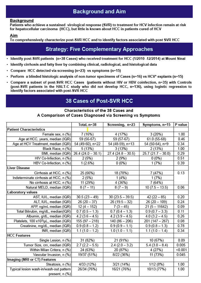
Background: Patients who achieve a sustained virological response (SVR) to treatment for chronic hepatitis C (HCV) remain at risk for hepatocellular carcinoma (HCC). Aims: To characterize patients who developed HCC more than 12 mo after achieving SVR.
Methods: Demographic, laboratory, and HCC stage and treatment data on 41 cases were obtained from medical records (1/2010-12/2014). Histology of H&E stained non-tumor tissue was evaluated by blinded review using the Knodell system for necroinflammation (HAI scale, 0-18) and the Scheuer system for stage (scale, 0-4); the specimens were from 17 patients treated with resection or liver transplantation. Patients with HCC detected through surveillance were compared to those diagnosed when symptomatic.
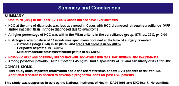
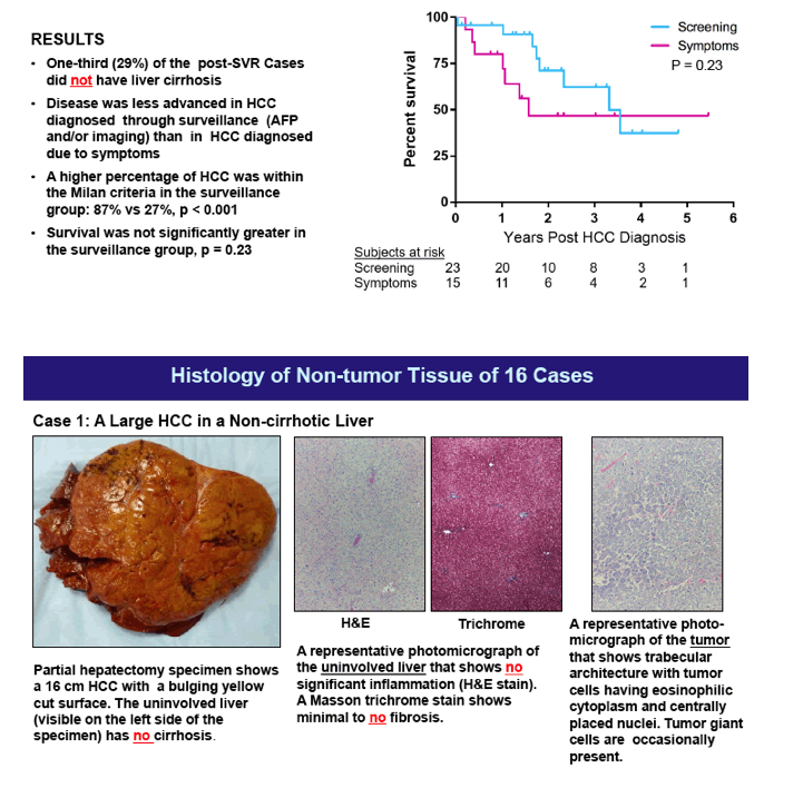
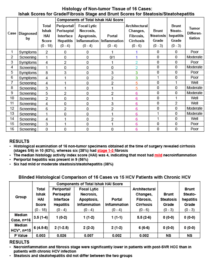
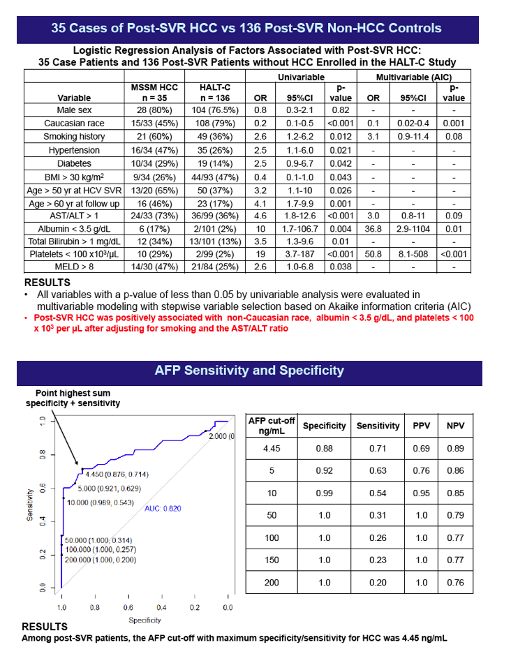
Results: HCC was diagnosed a median of 6 yr post-SVR when patients were a median age of 58 yr. Only 71% of the 41 cases had cirrhosis, and only 56% had AFP >10 ng/mL, 83% were male, and 88% were non-Asian. Comorbidities included diabetes (29%), HBV (2%), and HIV (12%). Median tumor size was 2.8 cm (range, 0.8-18.2), 83% had a single lesion on imaging and 51% had vascular invasion. Median laboratory values at the time of HCC diagnosis indicated that liver function was generally well-preserved: albumin 4.2 g/dL (2.1-5.0), platelets 148 x 103 cells/μL (48-446), and total bilirubin 1.3 mg/dL (0.2-8.8). HCC was diagnosed via surveillance (imaging and/or AFP) in 27 patients. Among the surveillance group, 85% were within Milan criteria versus 23% in the symptomatic group (p<0.01). Survival at 1 and 3 yr were 91% and 60% in the surveillance group and 62% and 62% in the symptomatic group (p=0.3). The histology study yielded important insights: Only 10/17 (59%) of the (non-tumor) tissues had stage 4 cirrhosis, whereas 6/17 (35%) had stage 0-2 fibrosis. Median HAI was 4 (range, 0-8), indicating that most had mild necroinflammation. Periportal hepatitis was present in 59%, only two had (mild) steatosis and three had steatohepatitis (5/17 = 29%).
Conclusions: Many patients lacked traditional HCC risk factors: 44% had AFP < 10 ng/mL and 40% of 17 histological specimens were non-cirrhotic and 70% lacked liver fat (steatosis/steatohepatitis). Future research should seek novel indicators of persistent HCC risk after HCV cure, e.g., somatic DNA mutations and epigenetic changes. This study did not show a survival benefit to screening; however, a higher percentage of patients diagnosed through surveillance were within Milan criteria.
|
| |
|
|
|
|
|