 |
|
|
| |
Exhausted T-cells and Inflammatory Monocytes
are Linked to Brain Atrophy in HIV
|
| |
| |
Reported by Jules Levn
CROI 2017 Feb 14-16 Seattle, WA
Brooks I. Mitchell1, Glen M. Chew1, Kalpana J. Kallianpur1, Mary Margaret Byron1, Robert H. Paul2, Beau K. Nakamoto1, Dominic C. Chow1,
Cecilia M. Shikuma1, Lishomwa C. Ndhlovu
1Hawaii Center for AIDS, University of Hawaii, Honolulu, Hawaii, USA. 2The Missouri Ins>tute of Mental Health, University of Missouri-St. Louis, St. Louis, Missouri, USA.
-------------------------------
CD8-Cell Exhaustions Predicts Primary HIV Infection Progression
HIV Causes CD8 Exhaustion-Occurs Early/Predicts HIV
Progression.....http://www.natap.org/2016/IAC/IAC_90.htm
Changes in markers of T cell senescence and exhaustion with Atazanavir-, Raltegravir-, Darunavir-Based Initial Antiviral Therapy: ACTG 5260s......"Overall, sustained declines over time were evident in all treatment groups for CD4+ T cell markers of senescence and exhaustion.....HIV-infected patients with low-level CD4+ T cell count on ART have more senescent CD4+ T cells compared to those with increased CD4+ T cell levels.....these results add to the current literature outlining the incomplete reversal of inflammation, senescence and immune activation in the setting of effective treatment"....The age-associated remodeling of the immune system, through accumulation of senescent T cells may contribute to numerous aging-related pathologies [2]. Senescent T cells have been associated with CVD.....We did not find any benefit of RAL over PIs on reducing immunosenescence or exhaustion during the first 96 weeks of successful treatment......Sustained decreases (about 31% or more) from baseline in levels of % CD28-CD57+ of CD4+ T cells were evident among all treatment groups (Figure 1, Table 1). In contrast, there was no apparent change from baseline to 96 weeks in levels of % CD28-CD57+ of CD8+ T cells. For exhaustion markers, declines from baseline were evident for % PD1+ of both CD4+ and CD8+ T cells across all treatment groups, by week 24, with further apparent declines for CD4 + T cell exhaustion by week 96; week 96 levels were on average at least 56% lower than baseline levels. Treatment group differences were not apparent for any immunosenescence and exhaustion marker (p>0.1;Supplemental Table 1). Overall, sustained declines over time were evident in all treatment groups for CD4+ T cell markers of senescence and exhaustion......At baseline, plasma viral load was correlated with markers of exhaustion (PD1+) (r=0.24-0.31; p<0.001), but not senescence (|r|<0.15, p>0.05). With regards to associations of baseline markers of immunosenescence with markers of T-cell and monocyte activation, or inflammation at baseline (Supplemental Table 2), the strongest associations were apparent between % of CD4+ CD38+DR+ and % of CD4+ CD28-CD57+ (r=0.42) and % of CD4+ PD1+(r=0.40) (Supplemental Figure 2). There was also a moderate association at baseline between monocyte activation (sCD163) and CD4+ exhaustion (% of CD4+ PD1+) (r=0.35). These moderate associations were also apparent when examining the concurrent changes over 96 weeks between these set of markers with correlations ranging from 0.33 to 0.55 (Supplemental Table 3); in contrast, a moderate association between the levels of these markers at week 96 was only apparent for CD4+ activation and senescence (r=0.44)" http://www.natap.org/2016/HIV/070616_02.htm
"PD-1 blockade has resulted in improved virologic outcomes in models of chronic HIV-1 infection....."It is possible that a treatment regimen involving inhibitory receptor blockade could result in durable ART-free virologic control, but the only way to test this possibility is by proceeding with carefully designed human clinical trials that assess the safety and efficacy of inhibitory receptor blockade in individuals on effective ART.....http://www.natap.org/2015/HIV/100715_02.htm
------------------------------------------------
Abstract Body:
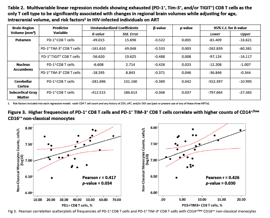
We have previously reported that T-cell exhaustion associated with neurocognitive impairment among HIV-infected adults on stable antiretroviral therapy (ART). Here, we investigated the relationship between immune populations and regional brain volumes.
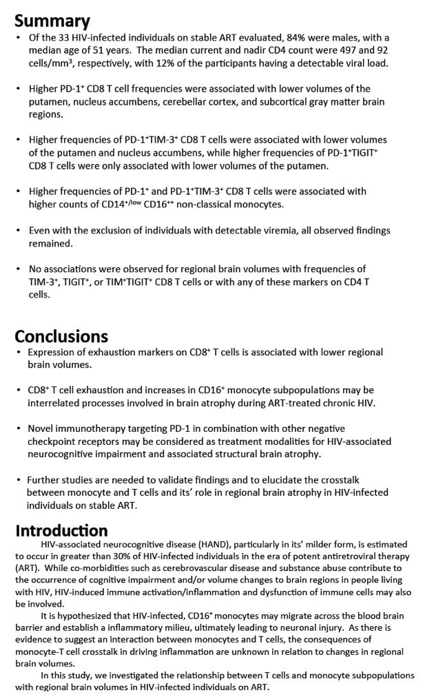
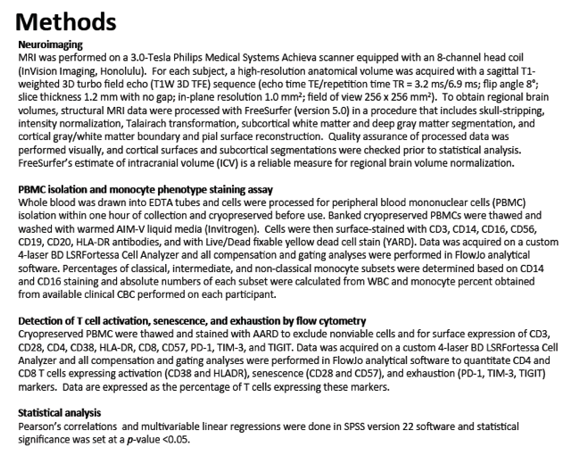
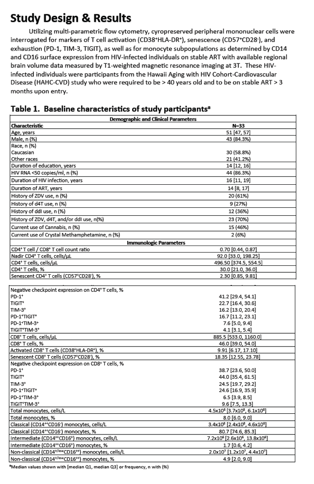
We utilized flow-cytometry to assess markers of CD8 T-cell activation (CD38+HLA-DR+), senescence (CD57+CD28-), and exhaustion (TIM-3, PD-1 and TIGIT) in peripheral blood mononuclear cells in HIV-infected individuals on stable ART with available regional brain volume data measured by T1-weighted magnetic resonance imaging at 3T as well as monocyte counts for classical (CD14+CD16-), intermediate (CD14+CD16+) and non-classical/inflammatory (CD14+/lowCD16++) subsets. Multivariable linear regression was utilized to assess the relationships between the immunophenotypes and regional brain volumes while controlling for intracranial volume and age. Pearson correlations were used in the analysis.
Thirty-three HIV+ participants were mostly male (84%) with a median age 52 years, and median current CD4 T-cell count of 479 cells/mm3. Detectable viral load (>50 copies/ml) was noted in 12% of the participants. Higher PD-1+ CD8 T-cell frequencies (%) were associated with lower volumes in the putamen (β= -0.522, p=0.005), nucleus accumbens (β= -0.426, p=0.023), cerebellar cortex (β= -0.389, p=0.042), and subcortical gray matter (β= -0.368, p=0.037) brain regions. Higher % of PD-1+TIM-3+ CD8 T-cells were associated with lower volumes in the putamen (β= -0.533, p=0.003) and nucleus accumbens (β= -0.371, p=0.046), while higher % of PD-1+TIGIT+ CD8 T-cells were only associated with lower volumes in the putamen (β= -0.488,p=0.008). Higher % of PD-1+, PD-1+TIM-3+ and PD-1+TIGIT+ CD8 T-cells were associated with higher counts of inflammatory monocytes (all p<0.05). Even with the exclusion of individuals with detectable viremia, all findings remained significant. No associations were observed for regional brain volumes with % of TIM-3+, TIGIT+, or TIM+TIGIT+ CD8 T-cells or with any of these markers on CD4 T-cells.
T-cell exhaustion was associated with lower brain volumes and increases in inflammatory monocytes, suggesting monocyte-T-cell exhaustion may be interrelated processes involved in brain atrophy during ART-treated HIV. Immunotherapy targeting PD-1 in combination with other negative checkpoint receptors may be considered as treatment modalities for HAND and associated structural brain atrophy.
|
| |
|
|
|
|
|