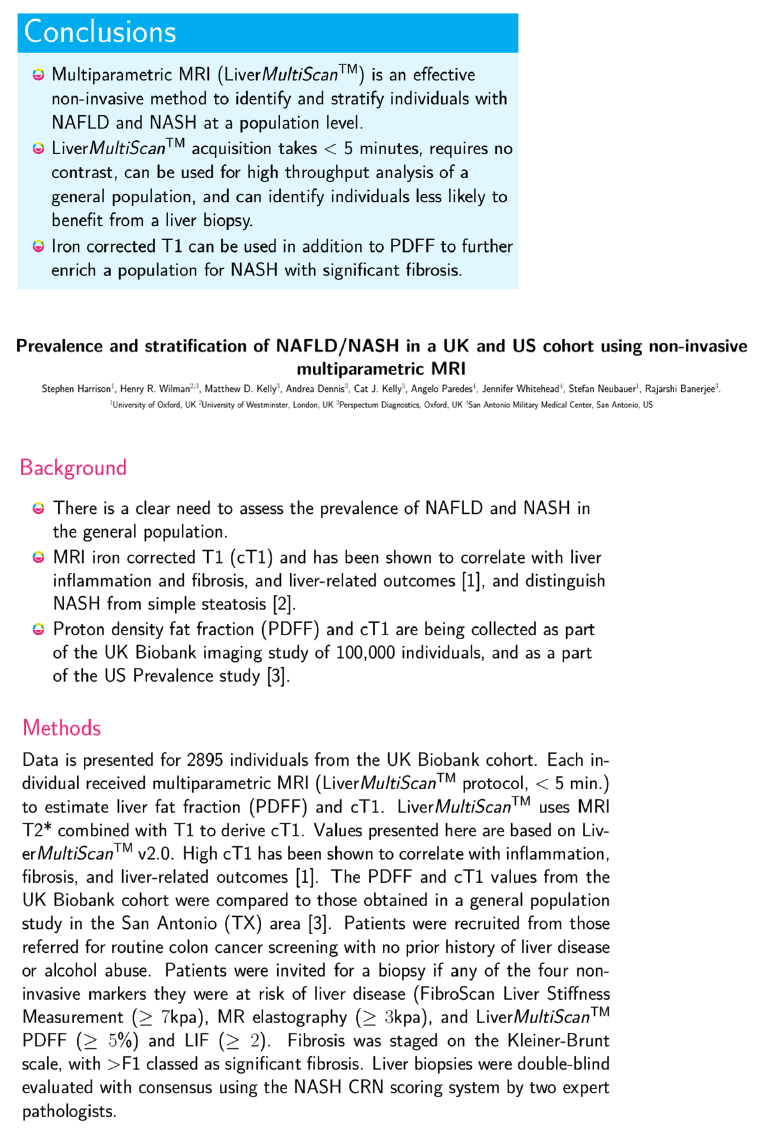
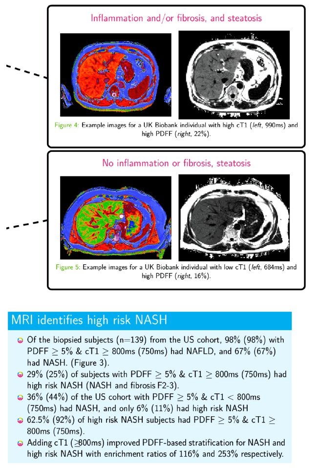
 |
|
|
| |
Prevalence and stratification of NAFLD/NASH in a UK and US cohort using non-invasive multiparametric MRI
|
| |
| |
Poster pdf attached
Download the PDF here
Dr. Stephen Harrison
Reported by Jules Levin
EASL 2018 April 11-15 Paris France
Program Abstract:
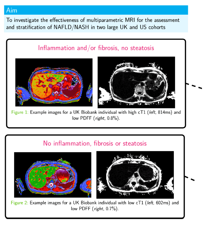
Background and aims
This is an ongoing prospective study with the following aims: 1) assess the prevalence and severity of NAFLD/NASH in San Antonio; 2) to evaluate the diagnostic performance of FibroScan-(FS) and magnetic resonance imaging (MRI)-based techniques in predicting the degree of hepatic steatosis, inflammation and fibrosis compared to liver biopsy (LB).
Methods
Patients referred for a routine colon cancer screening with no prior history of liver disease or alcohol abuse offered participation in the study. Screening for NAFLD using 5 modalities was performed. 2 FibroScan® (FS) modalities: liver stiffness measurement (FS-LSM) and Controlled Attenuation Parameter (FS-CAP) and 3 MRI-based imaging-based modalities: MR elastography (MRE-LSM), proton density fat fraction (MRI-PDFF) and liver inflammation fibrosis score (LMS-LIF, Liver Multiscan®). Patients with FS-LSM ≥7 kPa, MRE-LSM ≥3 kPa, PDFF ≥5%, or LIF ≥ 2 were proposed a LB. LB were assessed by two expert pathologists in a double-blind manner with consensus, using the NASH CRN scoring system. The FLIP algorithm was also utilized to assess degree of disease activity. Diagnostic performance was assessed using area under receiver operating curve (AUROC).
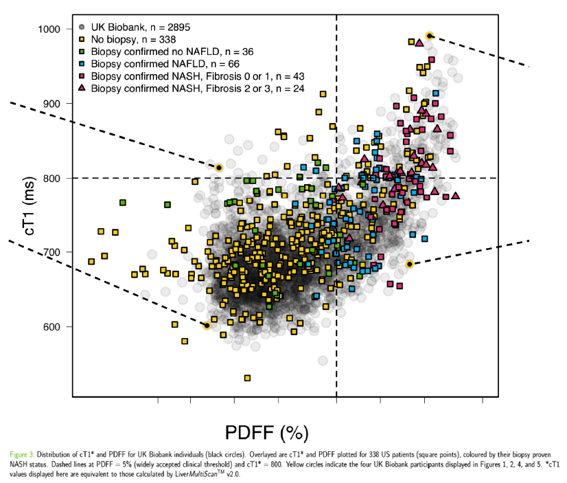
Results
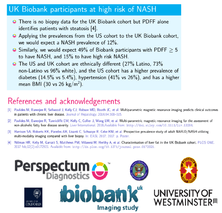
To date, 673 patients have been screened, of which 176 underwent LB. All imaging modalities performed in 449 patients. 328 patients (73%) had a normal liver (289 with normal imaging and 39 with normal LB). 63 (14%) and 58 (13%) patients had biopsy-confirmed NAFLD and NASH respectively. The prevalence of NAFLD and NASH was therefore 27% and 13%, respectively. Sex, BMI, ethnicity, diabetes status, hypertension status and lifestyle factors all were significantly linked to the normal/NAFL/NASH status. 160 patients were fully assessed (imaging modalities and LB): 24% were S0, 36% S1, 24% S2 and 16% S3. Respective AUROCs for FS-CAP and MRI-PDFF were for S ≥1: 0.80 [0.73; 0.88] and 0.93 [0.88; 0.98], for S ≥2: 0.82 [0.75; 0.88] and 0.96 [0.93; 0.99] and for S ≥3: 0.76 [0.68; 0.85] and 0.94 [0.89; 0.98]. 59% were F0, 28% F1, 9% F2 and 4% F3/F4. Respective AUROCs for FS-LSM, and MRE-LSM were for F ≥2: 0.81 [0.73; 0.89], 0.83 [0.73; 0.92].
Conclusions
Using several novel FS or MRI-based imaging markers, this study confirms the high prevalence of NAFLD and NASH among adults in south Texas. FS-CAP and MRI-PDFF are very good imaging modalities to non-invasively assess liver fat and FS-LSM/MRE are very good imaging modalities to non-invasively assess fibrosis in NAFLD.
|
| |
|
|
|
|
|