 |
|
|
| |
Retinal Thinning in Well-Controlled HIV-infected Patients
|
| |
| |
CROI 2020
Reported by Jules Levin
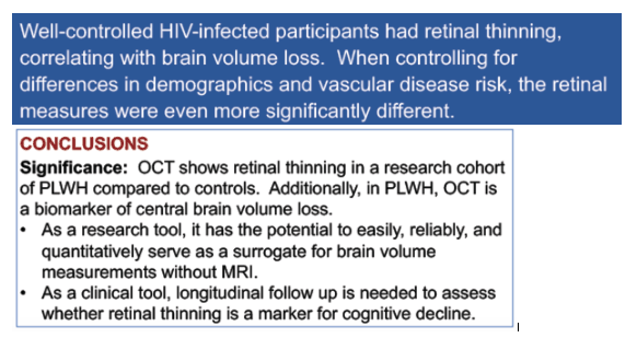
"PLWH on ART had thinning of the RNFL and the GC layer of the retina. This retinal thinning was asymptomatic but was strongly associated with measures of brain atrophy. This suggests that there is widespread neurodegeneration including the retina despite adequate ART."
Bryan Smith1, Katrina Geannopoulos1, Ramiro Maldonado2, Tianxia Wu1, Elizabeth F. Horne1, Lillian Ham3, Joseph Snow3, Govind Nair1, Daniel S. Reich1, Chuen-Yen Lau3, Emily Chew2, Avindra Nath1
1National Institute of Neurologic Disease and Stroke, 2National Eye Institute
3National Institute of Mental Health, 4National Institute of Allergy and Infectious Diseases National Institutes of Health, Bethesda, MD
Program Abstract
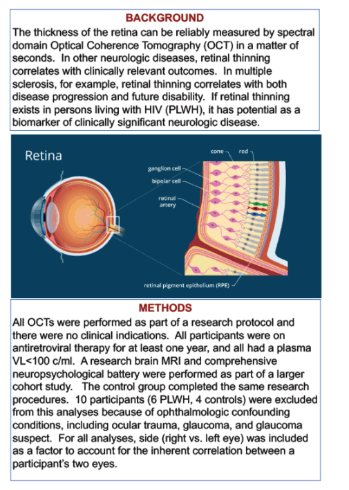
Retinal measurements correlate well with neurologic disease in multiple sclerosis, however whether such measurements correlate with neurologic disease in well-treated persons living with HIV (PLWH) is unknown. We evaluated differences in retinal measures by spectral domain optical coherence tomography (SD-OCT) between PLWH and uninfected controls and correlations with the retinal measures and brain volumes, neuropsychological (NP) function, and markers of neuronal injury and neuroinflammation.
SD-OCT was performed on 69 PLWH on antiretroviral therapy (ART) and 28 uninfected controls. Participants also underwent brain MRI, neuropsychological testing, and an optional lumbar puncture. All procedures, including the SD-OCT, were completed for research only and there were no clinical indications. Mean retinal nerve fiber layer (RNFL) and ganglion cell inner plexiform layer (GC-IPL) thicknesses were compared between groups using ANCOVA, and means were correlated with pre-selected MRI brain volumes, NP domain scores, and CSF cytokines and neurofilament light chain.
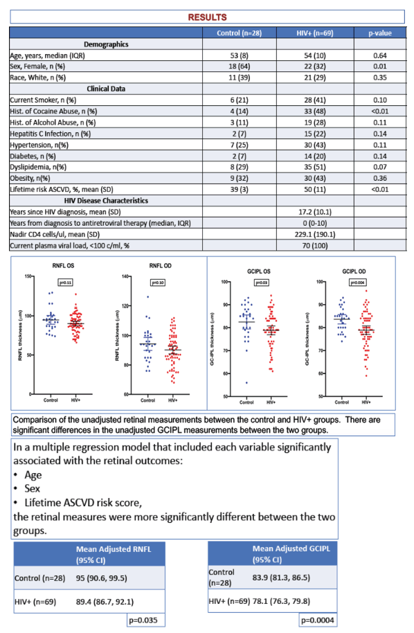
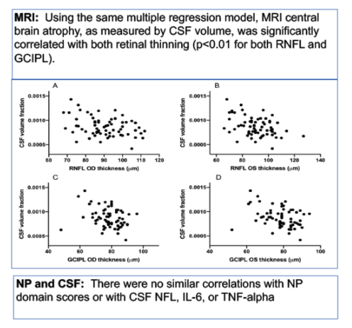
There were no differences in age, race or visual acuity between the two groups; there were more women in the control group (p=0.006). In the HIV+ group, the median time since diagnosis was 19 years and all had an HIV RNA level <100 copies/ml for at least one year prior to the SD-OCT. Multiple regression analyses indicated that the HIV+ group had thinner adjusted-mean RNFL (78.17m, 95% CI 76.3, 80.0; control = 84.0m, 95% CI 81.3, 86.5; p < 0.005) and GC-IPL (90.0m, 95% CI 87.0, 92.6; control = 96.6m, 95% CI 92.2, 101.0; p = 0.01). In the HIV+ group, retinal thicknesses were negatively associated with the fraction of CSF volume (i.e. brain atrophy) on MRI (p=0.01 for RNFL and 0.006 for GC-IPL). There were few associations with NP domains and CSF measurements.
PLWH on ART had thinning of the RNFL and the GC layer of the retina. This retinal thinning was asymptomatic but was strongly associated with measures of brain atrophy. This suggests that there is widespread neurodegeneration including the retina despite adequate ART.
|
| |
|
|
|
|
|