 |
|
|
| |
Mitochondrial disorders in activated memory CD4+ T cells derived from HIV-infected immunological non-responders
|
| |
| |
IAS 2021 July 18-22
Presenter: Violetta Vlasova
Authors: V. Vlasova * (1 ), E. Saidakova (1,2), L. Korolevskaya (1 ), N. Shmagel (3), S. Younes (4), M. Lederman (5), K. Shmagel (1)
Institutions: (1) Institute of Ecology and Genetics of Microorganisms (UB RAS), Perm Federal Research Center (UB RAS), Laboratory of Ecological Immunology, Perm, Russian Federation, (2) Perm State University, Department of Biology, Perm, Russian Federation, (3) Perm Regional Center for Protection against AIDS and Infectious Diseases, Medical Assistance Department, Perm, Russian Federation, (4) The Emory University School of Medicine, Department of Pathology & Laboratory Medicine, Atlanta, United States, (5) Case Western Reserve University School of Medicine, Cleveland, United States
Abstract:
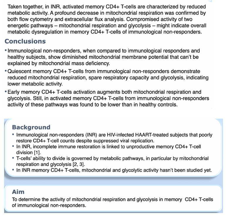
BACKGROUND: Immunological non-responders (INRs) are HIV-infected HAART-treated subjects that poorly restore CD4+ T-cell counts despite suppressed viral replication. In INRs, CD4+ memory T-cells cycle but don't divide, which is linked to chronic lymphopenia formation and adverse clinical outcome. As mitochondria impacts cell proliferation, we evaluated mitochondrial parameters in resting and stimulated CD4+ memory T-cells of IN Rs in vitro.
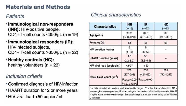
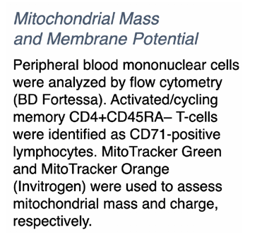
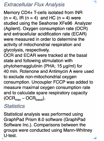
METHODS: Blood mononuclear leucocytes derived from IN Rs (CD4+ T-cells <350/ul), immunological responders (IR; CD4+ T-cells >500/ul) and healthy controls (HC) were analyzed by flow cytometry (BD Fortessa). Activated/cycling CD4+ memory T-cells (CD4+ TM; CD4+CD45RA') were identified as being positive for CD71. MitoTracker Green and MitoTracker Orange (lnvitrogen) were used to assess mitochondrial mass and charge, respectively. Oxygen consumption rate (OCR) was measured in isolated CD4+ TM using the Seahorse XFe96 Analyzer in order to determine mitochondrial respiration at basal state, after 40 minutes exposure to phytohemagglutinin (PHA; 15 ug/ml), and subsequent Rotenone/Antimycin A (0,5 uM) injection.
RESULTS: Mitochondrial masses were similar in CD4+ TM of HIV-infected and healthy subjects (p>0.05). However, mitochondrial charge in CD71 +CD4+ TM of IN Rs when compared to I Rs was decreased (p<0.05) indicating lower oxidative phosphorylation activity. In line with that, CD4+ TM derived from INRs compared with those derived from IRs (p=0.010) and HCs (p=0.0007) exhibited lower OCR. Subsequent stimulation of CD4+ TM with PHA increased OCR in all groups. Nevertheless, as opposed to activated cells of HCs, stimulated CD4+TM of HIV-infected subjects showed lower OCR (INR ' p<0.0001; IR ' p=0.028). Moreover in INRs, OCR in activated CD4+ TM was even lower than that in I Rs (p=0.027).
CONCLUSIONS: Our novel findings demonstrate that in immunological non-responders, CD4+ memory T-cells exhibit reduced mitochondrial respiration. This malfunction was shown in both resting and activated lymphocytes, and it can't be explained by mitochondrial mass deficiency. CD4+ memory T-cells are known to be the main source for immune reconstruction in HIV-infected patients receiving HAART. Thus, mitochondrial disorders found in these cells might be linked to poor immune restoration.
|
| |
|
|
|
|
|