| |
Stiffness and Impedance: The New Liver Biomarkers Editorial
|
| |
| |
Clinical Gastroenterology and Hepatology
Volume 5, Issue 10, Pages 1144-1146 (October 2007)
Raza Malik, MD1, Nezam Afdhal, MD2
Liver Center, Department of Medicine, Beth Israel Deaconess Medical Center; Harvard Medical School, Boston, Massachusetts
Dr Raza Malik was supported by a Liver Institute for Education & Research (LIFER) award and a St John Ambulance Air Wing Traveling Fellowship.
Dr Nezam Afdhal had received grant support from EchoSens and is the FDA sponsor of the registration trials for FibroScan. He has received research and consultancy support from Prometheus and Quest Diagnostics.
The universal end point of all chronic liver diseases is progressive hepatic fibrosis leading to architectural distortion and cirrhosis.1, 2 In patients with no clinical features of cirrhosis or portal hypertension, the diagnosis of compensated cirrhosis is important to establish, because these patients require screening for varices and hepatoma.3, 4 The liver biopsy has been pivotal to the diagnosis of cirrhosis since its advent more than half a century ago by Giorgio Menghini at the Mount Sinai Hospital and has become the established gold standard for the diagnosis of liver fibrosis. Why then has there been a logarithmic explosion of modalities appearing on the scene during the last decade to join or displace the percutaneous liver biopsy as the currently accepted gold standard? The most important limitation of liver biopsy is the small but significant morbidity and mortality associated with the procedure, which are probably underreported in clinical practice.5 In addition, the invasive nature of the biopsy creates great apprehension among patients, leaving them with an exaggerated view of potential complications. The liver biopsy is prone to sampling error because only 1:50,000 of the organ is sampled, with individual patients exhibiting great variation in stages of liver fibrosis. In fact, sampling error accounts for a 15% diagnostic inaccuracy between cirrhosis and Metavir stage 3 in patients with chronic hepatitis C who had paired laparoscopic biopsies of both liver lobes.6 The size of the liver biopsy sample also appears to be critical, with a size of >2 cm required to make histologic assessment satisfactory, making technical expertise important, and even with an adequate biopsy size, histologic interpretation can be variable.7 Thus there is an important clinical role to be established for noninvasive modalities to screen for cirrhosis. A secondary clinical role for noninvasive tests has been to differentiate mild disease (Metavir fibrosis stage 0-1) from more advanced fibrosis (Metavir 2-4).
The current issue of Clinical Gastroenterology and Hepatology contains 3 articles on detection of liver fibrosis by using novel physical properties associated with progressive hepatic fibrosis.8, 9, 10 Hepatic elastography with magnetic resonance imaging (MRI) or ultrasound and splenic artery impedance with Doppler ultrasonography are all performed with a high degree of accuracy in diagnosing cirrhosis and represent new advances in imaging the physical properties of the liver and portal vasculature.
Elastography is a relatively simple evaluation of the liver stiffness and is based on the finding that as the liver becomes fibrotic, it becomes harder and less elastic. Ultrasonic elastography is in widespread clinical use in Europe and undergoing Food and Drug Administration registration studies in the United States. A dedicated ultrasonic system known as the FibroScan (Echosens, Paris, France) has been widely studied,8 and Talwalker et al have analyzed the reported literature in a meta-analysis to evaluate the clinical utility of the device for liver fibrosis. They showed excellent results for estimating cirrhosis, with sensitivity and specificity of 87% and 91%, respectively, from 9 pooled studies. The results for differentiating mild from advanced fibrosis were less robust (Table 1), and the issue might be the lack of uniformity of stiffness cutoffs between the various studies, making interpretation between studies difficult. The advantages of transient elastography with the FibroScan device are the ease of use, reproducibility, patient acceptance, and ability to integrate the device into an outpatient hepatology clinic. The disadvantages of transient elastography are that it might be unreliable in morbid obesity, it requires an acoustic window, and it only scans 1:100 of the liver and is therefore still susceptible to sampling error.
Table 1. Diagnostic Accuracy of MR Elastography, Ultrasonic (US) Elastography, and Splenic Doppler at Identifying Cirrhosis (F4) and Advanced Fibrosis (F2/F3/F4)
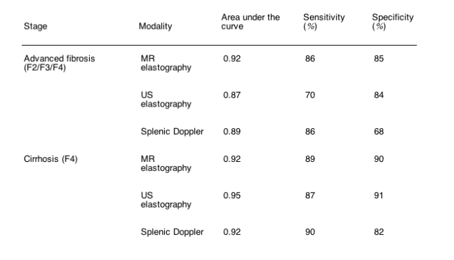
Yin et al9 investigated the use of MR elastography in assessing hepatic fibrosis in patients with chronic liver disease from various etiologies in a small pilot study (9). The technical issues with MR elastography are significant but seem to have been resolved so that elastograms from a large cross-sectional area of the liver can be obtained without motion artifact, and tissue shear stiffness can be calculated in kilopascals. The overall stiffness by MRI is considerably less than reported by ultrasonography in both normal subjects and patients, but it might reflect the different methodologies for calculating shear stiffness. Overall, MR elastography was able to identify cirrhosis and discriminate between moderate/severe fibrosis (F2-F4) and mild fibrosis (F1) (Table 1). Interestingly, there was a difference in shear stiffness between normal controls and stage 0 patients, and this will require further evaluation because the degree of fibrosis cannot explain this anomaly. The differences in stiffness were less significant between different fibrosis stages, but this study clearly shows the potential for MR elastography as a diagnostic tool.
The advantages of MR elastography are the potential to scan the whole liver and avoid sample bias, the lack of need for an acoustic window, and the ability to simply add elastography onto a standard MR exam of the liver. However, the inconvenience and expense of MR-based elastography compared with ultrasound are significant, and it is highly unlikely that MR elastography will be either cost-effective or have a high patient acceptability as a screening test for liver fibrosis. Patients who are undergoing liver MRI for other indications might represent a subpopulation in whom MR elastography can be performed. A head-to-head comparison of the 2 elastographic techniques would be of interest and could be combined with a cost-effectiveness analysis to identify best utilization strategy for stiffness testing.
In the final article, Liu et al10 use Doppler ultrasonography to assess hepatic fibrosis in patients with chronic hepatitis C infection. They performed a large, well-designed study consisting of both a training set and a validation set totaling 503 patients all undergoing liver biopsy. Duplex Doppler ultrasonography evaluated the splenic and hepatic arteries and the portal vein hemodynamics. The splenic Doppler impedance ratio was the major determinant of advanced fibrosis and cirrhosis, yielding excellent results with an area under the curve of 0.89 and 0.92, respectively (Table 1). However, there was a great variation in diagnostic accuracy (in terms of sensitivity and specificity) over a narrow range of Doppler indices, making this modality susceptible to operator-dependent inaccuracies. The ability to add these studies onto routine ultrasonography is clearly an advantage, but the issue of body habitus, effect of body mass index, and operator training and variability will need to be addressed.
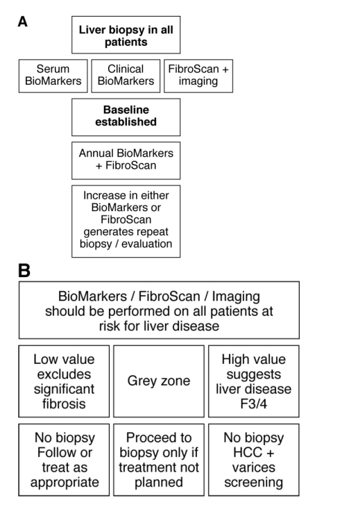
What do all these new tests mean for the clinician and the patient? They certainly offer excitement that we will have multiple options for noninvasively staging liver fibrosis and might reduce the need for liver biopsy to stage disease and to diagnose cirrhosis (Figure 1). The cross-sectional diagnosis of cirrhosis is just as effective now with serum tests and elastography as with liver biopsy.11, 12, 13 However, these noninvasive tests might also result in entirely new approaches to patients with liver disease. The ALT has long been the traditional screening test for liver disease, but now we might have an alternative rapid test in elastography and serum markers to identify the presence of fibrosis. Conceptually we could screen high-risk populations with a combination of ALT, serum biomarkers, and elastography and identify those with liver disease. The applicability of this approach to the large population of patients with nonalcoholic fatty liver disease and metabolic syndrome is particularly exciting if the issues of truncal obesity and an adequate acoustic window for liver stiffness measurement can be solved. The optimal utilization of these tests and their use in monitoring patients over time will require more clinical investigation, but these studies in Clinical Gastroenterology and Hepatology provide hope for the future development of this exciting field of new diagnostic algorithms for our patients with liver disease.
Figure 1. Potential diagnostic algorithms for utilization of noninvasive tests for liver fibrosis. (A) Combination pathway combining noninvasive tests with biopsy; (B) screening pathway using noninvasive tests before liver biopsy in at-risk patients.
References
1. 1Friedman SL, Rockey DC, Bissell DM. Hepatic fibrosis 2006: report of the Third AASLD Single Topic Conference. Hepatology. 2007;45:242-249. MEDLINE | CrossRef
2. 2Afdhal NH, Nunes D. Evaluation of liver fibrosis: a concise review. Am J Gastroenterol. 2004;99:1160-1174. MEDLINE | CrossRef
3. 3Jensen DM. Endoscopic screening for varices in cirrhosis: findings, implications, and outcomes. Gastroenterology. 2002;122:1620-1630. Abstract | Full Text | Full-Text PDF (214 KB) | MEDLINE | CrossRef
4. 4Chalasani N, Said A, Ness R, et al.. Screening for hepatocellular carcinoma in patients with cirrhosis in the United States: results of a national survey. Am J Gastroenterol. 1999;94:2224-2229. MEDLINE | CrossRef
5. 5Bravo AA, Sheth SG, Chopra S. Liver biopsy. N Engl J Med. 2001;344:495-500. MEDLINE | CrossRef
6. 6Regev A, Berho M, Jeffers LJ, et al.. Sampling error and intraobserver variation in liver biopsy in patients with chronic HCV infection. Am J Gastroenterol. 2002;97:2614-2618. MEDLINE | CrossRef
7. 7Bedossa P, Dargere D, Paradis V. Sampling variability of liver fibrosis in chronic hepatitis C. Hepatology. 2003;38:1449-145.
8. 8Talwalker JA, Kurtz DM, Schoenleber SJ, et al.. Ultrasound-based transient elastography for the detection of hepatic fibrosis: systematic review and meta-analysis. Clin Gastronerol Hepatol. 2007;5:1214-1220.
9. 9Yin M, Talwalker JA, Glaser KJ, et al.. A preliminary assessment of hepatic fibrosis with magnetic resonance elastography. Clin Gastroenterol Hepatol. 2007;5:1207-1213. Abstract | Full Text | Full-Text PDF (2339 KB)
10. 10Liu CH, Hsu S-J, Lin J-W, et al.. Noninvasive diagnosis of hepatic fibrosis in patients with chronic hepatitis C by Splenic Doppler impendance index. Clin Gastroenterol Hepatol. 2007;5:1199-1206. Abstract | Full Text | Full-Text PDF (734 KB)
11. 11Ziol M, Handra-Luca A, Kettaneh A, et al.. Noninvasive assessment of liver fibrosis by measurement of stiffness in patients with chronic hepatitis C. Hepatology. 2005;41:48-54. MEDLINE | CrossRef
12. 12Foucher J, Chanteloup E, Vergniol J, et al.. Diagnosis of cirrhosis by transient elastography (FibroScan): a prospective study. Gut. 2006;55:403-408. MEDLINE | CrossRef
13. 13Parkes J, Guha IN, Roderick P, et al.. Performance of serum marker panels for liver fibrosis in chronic hepatitis C. J Hepatol. 2006;44:462-474. Abstract | Full Text | Full-Text PDF (216 KB) | MEDLINE | CrossRef
|
|
| |
| |
|
|
|