| |
Fibrosis Progression Rates Double in Coinfected Within 2 Years
|
| |
| |
"Effect of hepatitis C virus (HCV) treatment in fibrosis progression rate (FPR) and time to cirrhosis (TTC) in patients co-infected with human immunodeficiency virus (HIV): A paired liver biopsy study"
Journal of Hepatology, Feb 2, 2007
Maribel Rodriguez-Torres et al. Fundacion de Investigacion de Diego, San Juan, Ave. De Diego 359 Suite 302, Santurce, PR 00909, Puerto Rico
Uncorrected Proof
NOTE from Jules Levin: this study examined several questions but a significant finding was the accelerated progression of fibrosis in coinfected patients, as the authos said: "....Of important clinical relevance, we showed that in an interval of time of 2 years, co-infected patients that are not treated for HCV have an injurious acceleration of FPR and substantial decrease in TTC....fibrosis progression rate doubled within the 2 years between biopsies...."
FPR: fibrosis progression rate; TTC: time to cirrhosis.
Paired (two) biopsies were perfomed. FPR of first biopsy (FPR1) was calculated as the ratio of staging of biopsy and duration of infection in years, and reported as Ishak fibrosis years (IshFy). Fibrosis progression rate for second biopsy (FPR2) was calculated as the ratio between the difference of second biopsy and the first biopsy staging, and the time interval between biopsies in years. Time to cirrhosis (TTC) was calculated by the ratio of first cirrhotic stage (Ishak staging 5) and FPR1 or FPR2. Patients were classified in three (3) groups according to IFN treatment received between biopsies: Group 1-no treatment, n=9; Group 2 IFN based therapy (IFN or IFN/RBV), n=30; Group 3-Peg-IFN alfa-2a based therapy (Peg-IFN-2a or Peg-IFN-2a/RBV) n=35. Liver biopsy in co-infected patients is repeated in non-cirrhotics at interval of time of more than 2 years, mainly for treatment consideration.
Fibrosis progression
FPR and calculated TTC at the time of both liver biopsies are shown in Fig. 1. TTC and FPR were not statistically different along the groups (p=0.91) at the time of first biopsy.
Fig. 1. Changes in fibrosis progression rate among treatment groups. Fibrosis progression rate (FPR): Group 1, No treatment, FPR1 0.22 (SD 0.17) Ish/y, FPR2 0.55 (SD 0.81) Ish/y, (p=0.37); Group 2, IFN, FPR1 0.26 (SD 0.34) Ish/y, FPR2 0.52 (SD 0.86) Ish/y, (p?0.001). Group 3, Peg-IFN Alfa-2a, FPR1 0.22 (SD 0.20) Ish/y, FPR2 -0.10 (SD 0.91) Ish/y, (p?0.001). ime to cirrhosis (TTC): Group 1, No treatment, TTC1 22.7 years, TTC2 9.09 years; Group 2, IFN, TTC1 19.23 years, TTC2 9.60 years; Group 3, Peg-IFN Alfa-2a, TTC1 22.7 years, TTC2 negative FPR (stabilization or regression).
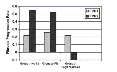
After the interval of time between biopsies, FPR2 in the no-treatment arm shows more than doubling of FPR, from 0.22 (SD 0.17) to 0.55 (SD 0.81), and TTC decreases from 22.7 years to 9.09 years. This difference in FPR in the non-treatment arm is not significant in this small cohort (p=0.37). A similar acceleration in FPR2 and TTC2 is seen in the group that received IFN based therapy (FPR2 0.52 vs. FPR1 0.26, and TTC, 19.2 years vs. TTC2 9.6 years, and was highly significant, p<0.001). The Peg-IFN based group had changes from FPR1 0.22 to a negative FPR2 -0.10 (p=0.001), , resulting in TTC1 22.7 years to TTC2 stable or regressive.
Discussion
The more rapid progression to cirrhosis and end stage liver disease in patients that are co-infected is well documented in the medical literature. However, factors that impact progression and the effect of HCV treatment in the liver disease are still not well understood. In a cohort of 274 HCV/HIV co-infected patients we demonstrated that HIV control, defined as undetectable HIV RNA level through HAART, resulted in slower FPR for patients at any HIV PCR level, and that HIV viral load, and not CD4+-independently predicted FPR [18]. It has been documented that alcohol use, gender, age at time of HCV infection, severe immune suppression, and ethnicity impact progression [15], [16], [17], [26].
In this study, we obtained a similar mean FPR before treatment in our co-infected patients to values reported in the literature [27]. The rationale to study progression by the calculation of FPR has been extensively published and utilized [28]. However, to study progression by use of liver biopsy histological interpretation and FPR has many limitations. Size and adequacy of biopsy, sampling issues, score and histopathologist expertise are confounder factors. We tried to decrease this variability by using a single experienced histopathologist, and assured all biopsies were longer than 25mm and had at least 12 portal spaces. Still, the calculation of FPR is an estimate, since it draws on the patient�fs accuracy of recollection of onset of risk behavior or event (first year of IDU or cocaine use for the majority of patients in this cohort). We performed two [2] separate structured interviews in order to decrease disparities of recollection but these cannot be controlled for all cases. FPR of the second biopsy or FPR2 is calculated by the interval of time between biopsies, and this measurement is more reliable than FPR1 dependent on estimate of time of infection.
In our cohort, we also attempted to eliminate as possible, confounding factors and differences among the groups, before and during the interval of time between biopsies, in order to study the effect of HCV treatment intervention. Our groups did not differ in the interval of time between biopsies nor the duration of HCV treatment. We assured no significant intervening alcohol use between biopsies and stable HIV characteristics. Moreover, baseline and virological characteristics of the treatment groups and control group were not different. Furthermore, we minimized selection for biopsy bias, as we perform liver biopsy in all patients that have no contraindication independent of estimated severity of liver disease.
In patients with chronic hepatitis C (CHC) without HIV co-infection, the beneficial effect of IFN on the natural fibrosis progression rate was found to be independent of genotype and early response [29]. Sobesky et al. demonstrated that median FPR decreased among all treated patients when compared to FPR before treatment. Furthermore, they demonstrated significant improvement in mean fibrosis stage among all treated patients, independently of response.
In HCV/HIV co-infected patients it has been reported that treatment with Peg-IFN alfa-2a as compared with IFN, significantly reduced fibrosis, in a standardized meta analysis study [22].
In the present study, we demonstrated that only patients that received Peg-IFN alfa-2a based therapy had improvements in grading and staging in the interval of time between biopsies, whereas patients that received no treatment or IFN based therapy had worsening of staging. When FPR and TTC were analyzed, patients that receive no HCV treatment have a rapid progression, with more than doubling of FPR in a mean interval of time of 2 years, decreasing the estimated TTC by more than half. Moreover, patients that received Peg-IFN alfa-2a based therapy had significant decreases in FPR2 and stabilization or regression of TTC2 and those that received IFN based therapy or no treatment had significant worsening and reduction of TTC2 by 60%.
Di Martino reported on the assessment of histological outcome in co-infected patients that received anti-HCV therapy with IFN, and confirmed that changes in histological response of >2 points decrease in total Knodell score, was achieved in 25% of all non-responders to treatment, independent of HIV status [30]. Improvements in fibrosis and overall HAI were also reported in patients treated with PEG-IFN/RBV in APRICOT, among patients that had bridging fibrosis or cirrhosis [23]. Of much interest were our findings when the results were analyzed among patients that achieved SVR or not with SOC Peg-IFN Alfa-2a/RBV. As expected, we had significant efficacy benefit of treatment with Peg-IFN/RBV. In this cohort, 44% SVR is similar to results of a global study in HIV/HCV (40%) [19].
In the mean interval of time after EOT, (around 12 months), there were no significant differences among patients that received a trial of treatment with Peg-IFN/RBV in the parameters of FPR2, or staging score between SVR and NR. This finding suggests that patients that did not achieve SVR obtained a beneficial stabilization of progression of fibrosis after HCV treatment intervention with Peg-IFN Alfa-2a/RBV. In order to examine the duration of the effect of Peg IFN alfa-2a/RBV in NR, studies with biopsies performed at longer intervals of time are required. However the deleterious effect in FPR and TTC in patients not treated or in those that received IFN based therapy will most probably worsen with longer intervals of time between biopsies.
To our knowledge our study is the largest cohort with paired biopsies in co-infected patients, to examine adequate duration IFN treatment effect on FPR and TTC. This study also adds to the body of evidence that Peg-IFN Alfa-2a/RBV therapy improves hepatic histology and suggests a decrease in progression to cirrhosis not only in SVR but also in NR. Our study has limitations, most importantly the reduced number of patients especially in the no treatment arm. This limitation will be difficult to correct even in prospective studies, because withholding HCV therapy in this population would be unethical in the majority of cases.
In summary, we have confirmed that HCV treatment intervention with Peg-IFN/RBV is beneficial in co-infection, not only to give patients best opportunity of SVR, but to achieve histological improvement, decrease progression of HCV related fibrosis and decrease risk to cirrhosis by stabilization or regression of TTC. Of important clinical relevance, we showed that in an interval of time of 2 years, co-infected patients that are not treated for HCV have an injurious acceleration of FPR and substantial decrease in TTC.
In this high-risk population with limited liver transplant opportunities and decreased efficacy with HCV treatment, benefits of treatment with Peg-IFN alfa-2a/RBV appear to go further than viral clearance. This is our only alternative at this point in time to decrease the high risk of liver related death in co-infected patients.
ABSTRACT
Introduction: Patients with hepatitis C (HCV) and human immunodeficiency syndrome (HIV) coinfection have rapid fibrosis progression. The effect on fibrosis progression rate (FPR) and time to cirrhosis (TTC) of HCV treatment has not been extensively studied.
Endpoints: 1 - Changes in FPR and TTC and staging after HCV therapy vs. no treatment 2 - Changes in FPR/staging of sustained viral responders (SVR) and non-responders (NR) to Peg-IFN alfa-2a and RBV.
Methods: Seventy-four (74) co-infected patients were grouped in three according to HCV treatment, Group 1 - None (n=9), Group 2 - IFN (n=30), Group 3-Peg-IFN alfa-2a (n=35). Paired liver biopsies were analyzed and FPR/TTC calculated for each biopsy.
Result: Baseline characteristics, duration of treatment and time between biopsies were similar among groups. HCV therapy, improved grading, but only Peg-IFN alfa-2a therapy resulted in staging decrease. Group 2 had significant staging increase and Group 1 had doubling of FPR2 and (TTC2) reduction from 22.7 to 9.09 years. Peg-IFN alfa-2a treated patients had negative change in FPR2 and stabilization in TTC. SVR and NR with Peg-IFN alfa-2a/RBV had same FPR2 and staging.
Conclusions: In patients with HIV/HCV co-infection Peg-IFN Alfa 2a based treatment produced regression or stable fibrosis in contrast to accelerated progression in those without treatment.
Introduction
Patients with chronic hepatitis C (HCV) and HIV co-infection have a more rapid progression toward chronic liver disease [1], [2], [3]. Chronic liver disease and end stage liver failure have become the most important cause for morbidity and mortality in co-infected patients [4], [5], [6], [7], [8], [9], [10], [11], [12].
Preliminary studies have demonstrated that HIV-HCV co-infected patients in highly active antiretroviral therapy (HAART) have slower fibrosis progression [13] and reduced liver related mortality [14]. Previous data support that age at HCV infection, daily alcohol intake, and necroinflamation score are independent predictors of increased fibrosis progression rate (FPR) [15], [16], [17]. We demonstrated that HIV viremia suppression is also associated to decreased fibrosis progression [18]. Strategies to decrease FPR include first of all, the attempt to achieve sustained viral response (SVR) through anti-HCV therapy. Co-infected patients have lower efficacy rates with standard of care (SOC) treatment for HCV than mono infected patients, achieving an overall sustained viral response (SVR) of 40% with better responses for genotypes 2 and 3 (62%) but significantly lower for genotype 1 (29%) [19], [20]. Treatment in HCV/HIV co-infected patients is challenging, because tolerability issues and medical contraindications are frequently encountered [21].
Treatment with IFN modalities, especially IFN/Ribavirin (RBV) and Peg-IFN/RBV, improves liver histology in HCV, even in patients do not achieve SVR [22]. There have been reports on the beneficial effect of Peg-IFN alfa-2a/RBV on histology of co-infected patients, showing benefits beyond viral clearance [23], [24]. However, the effect on FPR and the resultant outcome in time to cirrhosis (TTC) of HCV treatment has not been adequately studied. In this study, we compared the effect of HCV therapy intervention on FPR and TTC in a co-infected cohort with paired biopsies.
Methods
Patient selection
Patients with HCV/HIV co-infection that were evaluated at our site between January 1998 and December 2003 with paired biopsies were retrospectively examined. The clinical practice at our site is to obtain liver biopsies in all patients where the procedure is not contraindicated, as part of initial work up for therapy consideration. Liver biopsy is easily available and performed by invasive radiologist under CT guidance. Liver biopsy in co-infected patients is repeated in non-cirrhotics at interval of time of more than 2 years, mainly for treatment consideration. The main inclusion criterion for the study was the availability of paired biopsies with more than 12 portal spaces and 25mm of length. Patients included in this analysis had liver biopsies after 6 months of end of treatment (EOT). No patient in this study had second biopsy performed because evidence of hepatic tests elevation or suspicion of antiretroviral therapy (ART) hepatotoxicity. All liver biopsies were interpreted by the same experienced histopathologist (AF) using Ishak HAI grading 0-18 and staging 0-6 [25]. Patients were required to be naive to HCV therapy at the time of first liver biopsy and to have routine laboratory and viral parameters within 2 months of liver biopsy dates. During the period of time of the study we performed 248 liver biopsies in naive co-infected patients and 86 patients had paired liver biopsies. Patients with HBV co-infection, [2] active alcohol use before first biopsy or between biopsies [defined as more than 30g alcohol daily by structured interviews [3], or other causes of liver disease were excluded (Shistosoma mansoni) [1]. Patients that had changes in ART between biopsies [1] and had uncontrolled HIV viremia defined as >5000 HIV copies [2] or absolute total CD4<200 cells/mm3 at time of first biopsy were also excluded [1]. Patients were also required to have received at least 4 months of HCV therapy to participate (2 excluded). A total of 74 patients with paired biopsies were included for analysis in the study.
Data collection and analysis variables
Demographics and information regarding estimated time of HCV infection i.e. duration of time of main risk factor (blood transfusion before 1992, first use of injection drug use or cocaine use, (more than 80% of cohort), high risk sexual behavior as multiple partners or sex worker), to time of first biopsy, were obtained from structured interviews with the patients at 2 time points. HIV treatment and the average daily alcohol use for 5 years before the time of first biopsy, and between biopsies were obtained. FPR of first biopsy (FPR1) was calculated as the ratio of staging of biopsy and duration of infection in years, and reported as Ishak fibrosis years (IshFy). Fibrosis progression rate for second biopsy (FPR2) was calculated as the ratio between the difference of second biopsy and the first biopsy staging, and the time interval between biopsies in years. Time to cirrhosis (TTC) was calculated by the ratio of first cirrhotic stage (Ishak staging 5) and FPR1 or FPR2. Patients were classified in three (3) groups according to IFN treatment received between biopsies: Group 1-no treatment, n=9; Group 2 IFN based therapy (IFN or IFN/RBV), n=30; Group 3-Peg-IFN alfa-2a based therapy (Peg-IFN-2a or Peg-IFN-2a/RBV) n=35.
All patients signed informed consent document agreeing to the analysis of their confidential medical information for research purposes.
End points of study
The main end point of the study was to compare the effect among of IFN therapy in FPR and TTC as compared with a no treatment control group. The secondary end points were to examine effect on histology of IFN treatment in the interval of time between biopsies, and to examine staging and FPR among patients that achieved SVR or were non-responders (NR) of SOC treatment of Peg-IFN Alfa-2a/RBV, during the interval of time between biopsies.
Statistical analysis
Statistical analysis was performed using STATA (StataCorp, College Station, TX, v9.2). The Shapiro-Wilk test was used to test for normalcy of distribution of continuous variables. Since most of the variables were not normally distributed, we used the non-parametrical methods Wilcoxon matched-pairs signed-ranks, Mann-Whitney, and Kruskal-Wallis tests, when appropriate. The χ2 and Fisher�fs exact tests were used to compare categorical variables. A p value less than 0.05 was considered statistically significant.
Results
Baseline variables
The demographic and viral characteristics of the two [2] treatment groups and the no treatment group are shown in Table 1. There were no significant differences in any of the variables among treatment groups and the no treatment group. The majority of patients were male (75.7%), with mean duration of HCV infection of 20.0 years, and age at first biopsy of 43.6 years. HIV infection was fairly stable with a mean HIV log10 of 2.95 (SD 0.61) and absolute total CD4 cell counts of 524 (SD 292.00). Mean HCV log10 was 5.83 (SD 0.85) and most of the patients were genotype 1 (64.8%). Mean ALT levels were similar along groups (<3 times ULN), and most of the patients were using HAART (73%) vs. ART (20.5%) and only 6.5% were in no treatment.
Time between biopsies and duration of HCV treatment
Table 2 shows the interval of time between the [2] liver biopsies and the duration of HCV treatment. There are no differences between the interval of time between biopsies and the duration in months of HCV treatment among IFN and Peg INF alfa-2a based groups. HCV treatment mean duration was 9.60 (SD 5.64) months, (p=0.94), and mean time interval between biopsies was 1.95 (SD 0.50) years, (p=0.13). The mean duration of treatment for the IFN based group was 11.00 months and interval between biopsies of 2.11 years (range 1.25-4.00 years) for a mean interval of time after (EOT) of 1.19 years. The Peg IFN based group had a mean duration of treatment of 10.87 months, and mean interval of time between biopsies of 1.85 years (range 1.25-3.00 years) for a mean interval of time after EOT of 0.94 years.
Liver histology
Table 3 shows the histological findings of paired biopsies between treatment groups. Subjects from both treatment groups showed significant improvements in mean grading; IFN, -2.0 (SD 3.48, p=0.004), and Peg IFN alfa-2a -1.4 (p=0.004). The only group that had decreasing mean staging was Peg-IFN Alfa-2a based therapy [-0.06 (SD 1.64)] but the change did not achieve statistical significance (p=0.59). Patients that received IFN based therapy had significant worsening of staging in the interval of time of paired biopsies of +1.03 (SD 1.63), p=0.003. Patients that were not treated also had worsening of staging +0.77 (SD 1.20), but the change did not reach statistical significance (p=0.11).
Differences along SVR and NR
Table 4 shows patients that were treated with SOC Peg IFN Alfa-2a/RBV. As expected, best efficacy was obtained with SOC with 44% of patients achieving SVR.
When patients that achieve SVR are compared to those that are NR, there are no significant differences in FPR2 (-0.34 (SD 1.00) vs. 0.27 (SD 0.85), p=0.19), or along FS1 or FS2 of the SVR and NR (p=0.73 and 0.18) in the interval of time after EOT.
|
|
| |
| |
|
|
|