| |
HIV and Kidney Disease
|
| |
| |
(March 2008) Am Jnl of Kidney Diseases
"....Virus-specific kidney damage can be caused by direct infection of renal epithelial cells by HIV.....HIV was shown to infect epithelial cells of several segments of the nephron, including the glomerulus, tubules, and collecting duct. The pattern of epithelial-cell infection predicts the spectrum of histological abnormalities seen with HIVAN......Recent guidelines published by the Infectious Diseases Society of America to increase awareness of kidney disease and improve management recommend that all patients be assessed for existing kidney disease at the time of HIV diagnosis. These guidelines further recommend that patients with proteinuria of 1+ or greater on dipstick or eGFR less than 60 mL/min/1.73 m2 (<1 mL/s/1.73 m2) be referred to a nephrologist for further evaluation, including consideration of a kidney biopsy. Proteinuria assessment using quantitative methods should be encouraged to allow earlier identification of potential kidney disease.....the most important risk factors for CKD are undoubtedly hypertension and diabetes mellitus. Additional risk factors include a family history of kidney disease; African-American race; hepatitis C virus (HCV) coinfection; prior use of nephrotoxic medications, including some antiretroviral medications and antibiotics; and advanced HIV infection, reflected by high viral loads and low CD4 counts. Cocaine use, frequent in urban HIV-infected populations, has been linked to hypertensive renal changes in patients with HIV infection.....Because early and focused intervention is essential in treating and slowing the progression of kidney disease, accurate and efficient diagnosis is vital...
.....HCV-related kidney disease remains an important consideration in patients with HIV-HCV coinfection....Immune complex formation with HCV antigens can lead to HCV-associated glomerulonephritis with a membranoproliferative glomerular histopathologic state and/or cryoglobulin deposition.58 In addition to the presence of decreased complement levels and circulating cryoglobulins, the presentation of HCV in patients coinfected with HIV and HCV may include any combination of renal insufficiency, proteinuria, and hematuria....
.....Drug hypersensitivity reactions cause the vast majority of acute interstitial nephritis (AIN)....Numerous drugs are known to cause AIN. The HIV-infected population with exposure to a large number of medications may be at particular risk.37 In 1 biopsy study of patients with HIV infection, AIN was the fourth most common finding after HIVAN, hypertensive nephropathy, and FSGS. Frequent culprits included penicillin derivatives, cephalosporins, sulfa-containing drugs, quinolones, proton pump inhibitors, and NSAIDs (Table 4).36, 38 Time from drug exposure to evidence of AIN may vary from a few days, particularly with reexposure, to weeks or months. Prior tolerance of a drug does not rule it out as a current cause of interstitial nephritis....."
Kidney Biopsy in HIV: Beyond HIV-Associated Nephropathy
Am Jnl of Kidney Diseases
March 2008
Derek M. Fine, MD1, Mark A. Perazella, MD2, Gregory M. Lucas, MD, PhD1, Mohamed G. Atta, MD, MPH1
1 Johns Hopkins University School of Medicine, Baltimore, MD
2 Yale University School of Medicine, New Haven, CT
Case Presentation
A 57-year-old African-American woman with human immunodeficiency virus (HIV) infection for 18 years and hepatitis C presents with an acute decrease in glomerular filtration rate (GFR) and nephrotic-range proteinuria superimposed on chronic kidney disease (CKD). She was not on antiretroviral therapy because of nonadherence, and her viral load was 138,000 copies/mL, with a CD4 count of 365/μL. She had a remote history of intravenous cocaine and heroin abuse. Serum creatinine levels until 14 months earlier had ranged from 0.9 to 1.0 mg/dL (80 to 88.4 μmol/L; estimated GFR [eGFR] using the Modification of Diet in Renal Disease Study equation > 60 mL/min/1.73 m2 [>1 mL/s/1.73 m2]). One year before presentation, serum creatinine level was 1.2 mg/dL (106 μmol/L; eGFR, 58 mL/min/1.73 m2 [0.97 mL/s/1.73 m2]), and 9 months before, had increased to 1.9 mg/dL (168 μmol/L; eGFR, 34 mL/min/1.73 m2 [0.57 mL/s/1.73 m2]). At that time, a random urine protein-creatinine ratio was 10 g/g, and a 24-hour urine collection contained 11.3 g of protein. Urinalysis showed 10 white blood cells/high-power field, but no red blood cells. The patient refused a kidney biopsy and missed several appointments. She now presents with an increase in creatinine level to 3.2 mg/dL (283 μmol/L) and ongoing nephrotic-range proteinuria, with resolution of pyuria. She had used ibuprofen, 800 mg, several times weekly for several years to treat back pain. She has a 40-pack-year smoking history. On examination, her blood pressure was 140/92 mm Hg, pulse was 82 beats/min, and weight was 96 kg. Other than skin changes related to past drug use, physical examination findings were unremarkable. Laboratory results included white blood cell count of 4,800/μL, hemoglobin level of 10.9 g/dL, platelet count of 171 K/μL, normal complement levels, and negative antinuclear antibody test result. Ultrasound showed normal-sized kidneys with a slight increase in echogenicity. What clinical action is appropriate at this time?
Introduction
As we enter the second decade of highly active antiretroviral therapy (HAART), HIV-infected individuals are experiencing improved survival. Accompanying this is a new set of challenges unrelated to the immunosuppression of HIV infection. Included among these challenges is acute kidney disease and CKD. Despite a reduction in HIV-specific kidney disease in the HAART era,1 the HIV-infected population has both typical and disease-specific kidney disease risk factors that may result in different and varied disease presentation. Given the wide differential diagnosis in an HIV-infected patient with kidney disease, kidney biopsy is an essential tool for evaluation.
Increased Kidney Disease Despite Control of Virus-Related Kidney Disease
When assessing causes of kidney disease, it is important to distinguish between HIV-related and non-HIV-related disease. In recent years, morbidity and mortality from acquired immunodeficiency syndrome-related illnesses have decreased dramatically. However, rates of HIV-specific kidney diseases, the most common of which is HIV-associated nephropathy (HIVAN), have remained stable after an initial decrease in the mid-1990s.2 This initial decrease can be attributed to the widespread use of HAART.2, 3 Although HIVAN is still a major cause of end-stage renal disease (ESRD), ongoing use of HAART can prevent HIVAN,3 and prompt initiation of HAART can slow the progression of earlier stages of CKD caused by HIVAN.4, 5
Although the prevalence of HIVAN has not increased, that of non-HIV-related kidney disease in patients with HIV has.1, 6 In large part, this is related to an aging patient population, many of whom have diabetes mellitus and hypertension.7 In particular, African Americans are at increased risk of both diabetes mellitus and hypertension and also have a far greater incidence of ESRD than Caucasians (1,000 versus 259 per million, respectively).8, 9 In a high-risk urban HIV-infected African-American population in Baltimore, MD, with a mean follow-up of 10.6 years, an alarming 3.6% required renal replacement therapy, a rate that has not decreased during the HAART era.10 In a cross-sectional study of another urban HIV-infected population in New York City, 22% of African-American patients (versus 11.4% of whites) had either CKD or ESRD; 4.1% of the studied population including Caucasians and Hispanics had ESRD.11 Today, African Americans make up more than 50% of the new cases of HIV infection in the United States and 48% of the current HIV-infected population.12 This disproportionate representation in a population at particular risk of HIV-related and non-HIV-related kidney disease likely accounts for some of the increased prevalence in kidney disease witnessed in the entire HIV-infected population.
Identifying Kidney Disease in the HIV-Infected Population
Patients with HIV infection have other potential risk factors for kidney disease in addition to the more traditional kidney disease risk factors seen in the general population. In both patients with HIV infection and the general population, the most important risk factors for CKD are undoubtedly hypertension and diabetes mellitus. Additional risk factors include a family history of kidney disease; African-American race; hepatitis C virus (HCV) coinfection; prior use of nephrotoxic medications, including some antiretroviral medications and antibiotics; and advanced HIV infection, reflected by high viral loads and low CD4 counts.11, 13 Cocaine use, frequent in urban HIV-infected populations, has been linked to hypertensive renal changes in patients with HIV infection.14, 15
Because early and focused intervention is essential in treating and slowing the progression of kidney disease, accurate and efficient diagnosis is vital. Often, this is only possible with kidney biopsy. Recent guidelines published by the Infectious Diseases Society of America to increase awareness of kidney disease and improve management recommend that all patients be assessed for existing kidney disease at the time of HIV diagnosis. These guidelines further recommend that patients with proteinuria of 1+ or greater on dipstick or eGFR less than 60 mL/min/1.73 m2 (<1 mL/s/1.73 m2) be referred to a nephrologist for further evaluation, including consideration of a kidney biopsy.13 Proteinuria assessment using quantitative methods should be encouraged to allow earlier identification of potential kidney disease.16
Although some diagnoses can be presumed through clinical features and laboratory evaluations only, definitive diagnosis often will require kidney biopsy because many kidney diseases in patients with HIV infection have similar and overlapping presentations. Therefore, it is critical that HIV primary care providers and nephrologists understand the diseases affecting patients with HIV infection and recognize circumstances when biopsy is necessary.
Differential Diagnosis of Kidney Disease in Patients With HIV
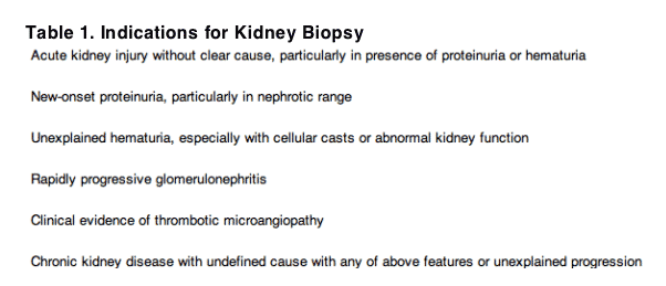
To determine the need for biopsy, it is essential to understand the clinical presentation and risk factors for each of the major kidney diseases affecting patients with HIV infection. Table 1 lists features of acute kidney disease and CKD that may indicate the need for kidney biopsy. Notably, the most common causes of acute kidney injury (AKI) in patients with HIV infection are decreased kidney perfusion and acute tubular necrosis caused by ischemia and/or medication exposure.17 In general, these entities are diagnosed by means of clinical and laboratory data.
HIV-1 Virus-Specific Glomerular Diseases
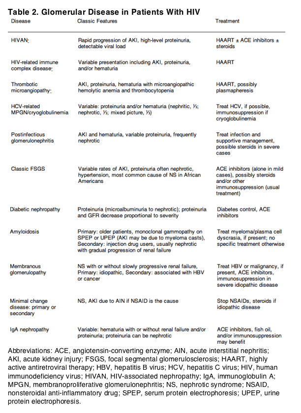
Virus-specific kidney damage can be caused by direct infection of renal epithelial cells by HIV, deposition of immune complexes composed of viral antigen-antibody complexes, and HIV-related thrombotic microangiopathy (TMA). The differential diagnosis of glomerular disease in this group includes many non-HIV-specific causes (Table 2) and is often difficult to distinguish on clinical grounds. Serological evaluation, including complement levels, hepatitis testing, and cryoglobulin levels, in addition to antinuclear antibody, antineutrophil cytoplasmic autoantibody, anti-glomerular basement membrane, and protein electrophoresis, if indicated, may suggest a diagnosis, but generally are not definitive.
HIV Associated Nephropathy
Abbreviations: ACE, angiotensin-converting enzyme; AIN, acute interstitial nephritis; AKI, acute kidney injury; FSGS, focal segmental glomerulosclerosis; HAART, highly active antiretroviral therapy; HBV, hepatitis B virus; HCV, hepatitis C virus; HIV, human immunodeficiency virus; HIVAN, HIV-associated nephropathy; IgA, immunoglobulin A; MPGN, membranoproliferative glomerulonephritis; NS, nephrotic syndrome; NSAID, nonsteroidal anti-inflammatory drug; SPEP, serum protein electrophoresis; UPEP, urine protein electrophoresis.
HIV-Associated Nephropathy
HIV was shown to infect epithelial cells of several segments of the nephron, including the glomerulus, tubules, and collecting duct. The pattern of epithelial-cell infection predicts the spectrum of histological abnormalities seen with HIVAN.18, 19 Histological features of HIVAN include collapsing focal segmental glomerulosclerosis (FSGS) as a result of podocyte proliferation20, 21 and tubular dilatation with atrophy and flattening of tubular epithelial cells.2, 21, 22 Interstitial edema and lymphocyte infiltration often accompany interstitial fibrosis in patients with HIVAN.
Definitive diagnosis of HIVAN is essential because untreated patients often will progress to ESRD within weeks to months.5 Treatment of patients with HIVAN consists of HAART, angiotensin-converting enzyme (ACE) inhibitors, and glucocorticoids.13, 22 HAART, in particular, was shown to slow the progression of CKD and increase dialysis-free survival in patients with HIVAN.4, 5
In the United States, HIVAN is exclusively an African-American disease.23 Clinically, the "classic" presentation of HIVAN is characterized by significant proteinuria (often protein > 3 g/d), serum creatinine levels greater than 2 mg/dL (>177 μmol/L), and progressive kidney failure.24 Edema and hypertension are uncommon in patients with HIVAN, and their absence may contribute to late recognition of kidney failure.24 Urinalysis often shows a bland sediment with varying numbers of proteinaceous casts and renal tubular epithelial cells.24
Notably, these clinical features are not specific to HIVAN and clinical presentation may vary; therefore, kidney biopsy is essential for early diagnosis of HIVAN. Nephrotic-range proteinuria has poor predictive value for HIVAN. In a study of 107 HIV-1-infected patients who underwent kidney biopsy, HIVAN was diagnosed in only 53% of the 55 patients with nephrotic-range proteinuria, yielding a sensitivity of only 73%.25 In patients with nephrotic-range proteinuria without HIVAN, common diagnoses included classic FSGS (21%), membranoproliferative glomerulonephritis (5%), amyloid A amyloidosis (4%), diabetic nephropathy (4%), and other diagnoses (12%). Additional analyses of this cohort showed that patients with nephrotic-range proteinuria and HIVAN had significantly lower CD4 T-cell counts compared with those with non-HIVAN renal disease. However, a low CD4 T-cell count (<200 cells/μL) in the presence of proteinuria also was not predictive of HIVAN because one third of these patients had kidney disease other than HIVAN. Another study of patients who underwent kidney biopsy and had recent measurements of HIV-1 RNA showed that viral load less than 400 copies/mL was associated with a low likelihood of HIVAN (1 of 23 patients). However, high viral load was not a sensitive indicator of HIVAN because patients with a viral load of 400 copies/mL or greater had HIVAN diagnosed in only 37% of cases (23 of 63 patients).1 Furthermore, the perception that large echogenic kidneys may aid in the diagnosis of HIVAN has no clinical foundation because most patients with HIVAN have normal-sized kidneys, and large kidneys may be seen with other pathological states.26 Therefore, although noninvasive indicators may be useful for excluding HIVAN, kidney biopsy is essential for its diagnosis.
HIV-Related Immune Complex Disease
Whereas HIVAN is a disease that predominately affects African Americans, immune complex disease directly related to the HIV infection is more prevalent in the Caucasian population.27 Patterns of glomerular involvement can be separated into 4 categories: immune complex-mediated glomerulonephritis, immunoglobulin A (IgA) nephritis, mixed sclerotic/inflammatory disease, and lupus-like disease.28
Immune complexes with HIV antigens were identified in both the circulation and kidney parenchyma of HIV-infected patients with IgA nephropathy and with immune complex glomerulonephritis.29, 30 Clinical signs of IgA nephropathy include hematuria and proteinuria that usually has protein less than 2 g/d.
Because these entities present similarly to other glomerular diseases and have no distinguishing clinical or laboratory findings, kidney biopsy showing the culprit lesion will significantly guide management, which, for these entities, includes HAART.
HIV-Related TMA
TMAs include thrombotic thrombocytopenic purpura and hemolytic uremic syndrome and are characterized by thrombocytopenia and microangiopathic hemolytic anemia. These disorders generally have kidney involvement, with AKI and variable levels of proteinuria and hematuria. TMA was described in patients with HIV infection and may resemble both thrombotic thrombocytopenic purpura and hemolytic uremic syndrome.31 Although the pathogenesis of HIV-related TMA is unknown, it is believed that damage to renal endothelial cells as a result of HIV infection is the primary event causing platelet activation and deposition in the microvasculature of the kidney.32
Cytomegalovirus infection also was implicated in the pathogenesis of TMA in this population.33 Similar to idiopathic TMA, plasma exchange therapy was used as primary treatment for patients with HIV-related TMA. Corticosteroids, splenectomy, immunoglobulin infusions, and antiplatelet agents also were used in patients with HIV-related TMA with variable degrees of success.32, 34 To decrease viral antigen levels, starting HAART would also be sensible.
The initial diagnosis of TMA in HIV-infected patients may be difficult because other clinical features typical of TMA, including thrombocytopenia, anemia, neurological dysfunction, nephropathy, and fever, are commonly seen in HIV-infected patients due to various other causes.34 Hence, biopsy is useful for a definitive diagnosis for any patient with proteinuria and AKI with systemic features of TMA.
Drug-Related Kidney Disease
Acute Interstitial Nephritis
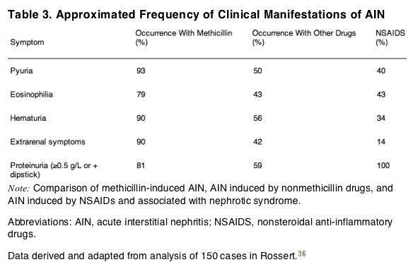
Drug hypersensitivity reactions cause the vast majority of acute interstitial nephritis (AIN). The seminal characterization of drug-induced AIN was derived from a report of 14 patients with methicillin-induced AIN. In these patients, increased serum creatinine levels were accompanied by rash, fever, and peripheral eosinophilia (the classic triad of AIN) 29%, 100%, and 100% of the time, respectively.35 In general, AIN from nonmethicillin drugs presents differently. Therefore, reliance on the "triad" to diagnose AIN may lead to misdiagnosis and underdiagnosis of AIN. The complete triad is seen in only 5% of patients with non-methicillin-induced AIN.36 The relationship between clinical findings and different forms of drug-induced AIN is listed in Table 3 and is based on approximately 150 case reports.36 Note that features including pyuria and eosinophilia, as well as extrarenal symptoms including fever, rash, or flank pain, are frequently absent in patients with non-methicillin-induced AIN. Therefore, the presence of kidney failure itself should raise concern for this entity even in the absence of clinical clues. It also is important to note that interstitial nephritis from nonsteroidal anti-inflammatory drugs (NSAIDs) usually has bland urinary sediment with the almost universal presence of proteinuria, occasionally in the nephrotic range (with an underlying minimal change or membranous lesion seen on histopathologic examination).
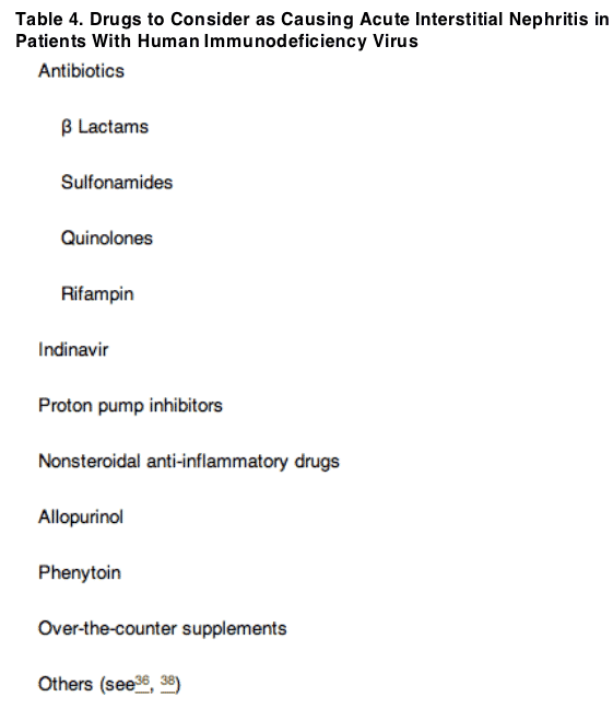
Numerous drugs are known to cause AIN. The HIV-infected population with exposure to a large number of medications may be at particular risk.37 In 1 biopsy study of patients with HIV infection, AIN was the fourth most common finding after HIVAN, hypertensive nephropathy, and FSGS. Frequent culprits included penicillin derivatives, cephalosporins, sulfa-containing drugs, quinolones, proton pump inhibitors, and NSAIDs (Table 4).36, 38 Time from drug exposure to evidence of AIN may vary from a few days, particularly with reexposure, to weeks or months. Prior tolerance of a drug does not rule it out as a current cause of interstitial nephritis.
In 1 series of 32 patients with AKI, only 44% with a clinical diagnosis of AIN had histopathologic evidence of this renal lesion.39Conversely, AIN is frequently found on biopsy when prebiopsy suspicion is low. Diagnosis is supported by recovery of kidney function with withdrawal of the suspected culprit drug. However, kidney biopsy showing active inflammatory interstitial infiltrates with or without interstitial eosinophils may be needed to appropriately clinch a diagnosis and guide management.
The most common treatment approach when AIN is suspected is discontinuation of any suspected drugs. This is a difficult and challenging task for patients on multiple medications that have the potential to promote AIN. Persistent kidney failure after drug discontinuation merits a kidney biopsy. Other than drug withdrawal, supportive care with dialysis, if needed, is the mainstay of therapy. Earlier discontinuation of an offending drug holds better hope for return to baseline kidney function because tubular atrophy and interstitial fibrosis can occur as early as 2 weeks after drug exposure.40
Although no controlled studies were performed regarding treatment of patients with AIN, corticosteroids (prednisone, 1 mg/kg, for 2 weeks with a taper) can be considered if kidney failure does not improve within a few days to a week after discontinuation of the offending drug.41Kidney biopsy may help guide this management decision.
Crystal Nephropathy
Various medications can crystallize in the renal tubular lumen, including indinavir, atazanavir, sulfadiazine, ciprofloxacin, and intravenous acyclovir, with precipitation of drug crystals occurring because of insolubility of the drug in urine. Volume depletion with sluggish urine flow rates is the most important risk factor for crystal nephropathy. Increased risk is also present in individuals with decreased GFR from underlying kidney disease; this enhanced risk may reflect excessive drug dosing, increased urinary concentrations of insoluble drug, and pH changes, all factors that may increase crystal precipitation.42 There generally is little role for kidney biopsy with these disorders because they can be diagnosed by means of clinical history of culprit drug exposure in the context of known risk factors accompanied by imaging studies, urine sediment examination for offending crystals, and stone analysis. Crystalluria is particularly common with indinavir use,43 but has become less common because use of this medication has decreased.
More recently, there were several case reports of nephrolithiasis caused by atazanavir. After the publication of 2 case reports in which atazanavir nephrolithiasis was confirmed by means of stone analysis,44, 45 Food and Drug Administration researchers reviewed the Adverse Event Reporting System database for similar cases.46 This showed 30 cases during a 4-year period, with 12 stones confirmed to be atazanavir by means of infrared spectrophotometry or other analysis. Additional studies estimated a prevalence of 0.97% of atazanavir stones in patients administered atazanavir. Although no associated risk factors were confirmed, atazanavir stones appear to form in alkaline urine.47 Although a relatively uncommon event, one must consider the possibility of atazanavir nephrolithiasis in patients administered atazanavir who develop renal colic. Finally, several cases of ciprofloxacin-associated crystal nephropathy were reported in non-HIV-infected patients.48 Because patients with HIV infection frequently are treated with this antibiotic, crystal nephropathy should be considered in the differential diagnosis of AKI in these patients. Patients with HIV infection with impaired kidney function, those who are volume depleted, and those with a urine pH greater than 6.0 are at greatest risk. Examination of urine sediment after drug exposure will facilitate diagnosis, potentially obviating the need for a kidney biopsy. To prevent AKI and crystalluria, ciprofloxacin should be dose adjusted for kidney function, patients should be volume replete, and alkalinization of urine should be avoided.
Tenofovir Toxicity
There is a growing body of evidence from retrospective case reports of changes in kidney function associated with the use of tenofovir, a nucleotide reverse-transcriptase inhibitor. Tenofovir is structurally similar to adefovir and cidofovir, both antiviral agents known to have nephrotoxicity. However, clinical trials involving tenofovir failed to show serious impairment of kidney function when evaluated by using eGFR or laboratory abnormalities in serum creatinine and serum phosphorus values.49, 50 Nevertheless, a retrospective analysis of a large observational cohort of patients administered either tenofovir (n = 344) or an alternative nucleotide reverse-transcriptase inhibitor (n = 314) as part of a HAART regimen showed that use of tenofovir was associated with a greater decrease in kidney function compared with use of other nucleotide reverse-transcriptase inhibitors.51 Patients who received tenofovir had a significantly greater decrease in estimated creatinine clearance that was noticeable after 90 days of initiating tenofovir treatment and persisted during the 1-year follow-up period. There was a 4% relative median decrease in GFR in patients who received tenofovir compared with those who received a different nucleotide reverse-transcriptase inhibitor, although both groups experienced GFR decrease.
No cases of AKI or Fanconi syndrome were reported in initial clinical trials of tenofovir; however, with its more widespread use in the general HIV-infected population, both AKI and Fanconi syndrome have been reported.52, 53, 54, 55, 56, 57 Of 27 patients with tenofovir-associated AKI recently reviewed by Zimmermann et al,57 16 (60%) had Fanconi syndrome. Fanconi syndrome resolved in all patients with discontinuation of tenofovir; however, 5 patients had incomplete recovery of kidney function after discontinuation of tenofovir (mean follow-up, 7.5 months; range, 3 to 20 months). It is this lack of full recovery in several case reports that highlights the need for early and accurate diagnosis of tenofovir toxicity. It is important to note that the high proportion of reported patients with Fanconi syndrome probably reflects publication bias, perhaps reflecting heightened awareness because of the known association of Fanconi syndrome with the parent drug adefovir. Accordingly, the presence of GFR decrease without Fanconi syndrome is the more frequent presentation of tenofovir nephrotoxicity.51
In the absence of other obvious explanations, one should assume that an isolated increase in creatinine level is related to tenofovir exposure. However, when there is a broader differential diagnosis, kidney biopsy may be helpful to evaluate for other potential causes of AKI. Because of diminished clearance of tenofovir in the context of substantial GFR decrease associated with AKI, the drug should be discontinued or dose reduced even if kidney disease is not caused by tenofovir. In the setting of tenofovir nephrotoxicity, kidney biopsy typically shows acute tubular necrosis without glomerular or interstitial changes.54, 57 If the kidney biopsy specimen shows an alternative explanation for kidney failure and kidney function recovers, tenofovir therapy often may be resumed. Close monitoring is required with dose reduction because kidney recovery may result in underdosing of tenofovir.
Other Non-HIV-Related Kidney Disease
Other diseases common in the general population should be considered in HIV-infected patients. These may have very similar or overlapping clinical presentations and therefore may be differentiated only by means of biopsy. Potential glomerular diseases include classic FSGS, the most common cause of nephrotic syndrome in African Americans, and IgA nephropathy, the most common cause of glomerulonephritis worldwide. Also of increasing importance are hypertensive nephrosclerosis and diabetic nephropathy.1 Other glomerular diseases that may require biopsy for diagnosis include amyloid A amyloidosis, lupus nephritis, membranous nephropathy, and postinfectious glomerulonephritis (Table 2).
Renal lymphoma and other infiltrative disorders also can be diagnosed by means of biopsy.
HCV-Related Kidney Disease
HCV-related kidney disease remains an important consideration in patients with HIV-HCV coinfection. The presence of HIV-HCV coinfection is particularly common in patients with a history of injection drug use. Immune complex formation with HCV antigens can lead to HCV-associated glomerulonephritis with a membranoproliferative glomerular histopathologic state and/or cryoglobulin deposition.58 In addition to the presence of decreased complement levels and circulating cryoglobulins, the presentation of HCV in patients coinfected with HIV and HCV may include any combination of renal insufficiency, proteinuria, and hematuria.58, 59 Treatment of patients with HCV, usually with interferon-based regimens, would be recommended in those with clinically significant kidney disease. Because interferon-based treatment has significant toxicity, kidney biopsy is important to confirm this diagnosis.
Postinfectious Glomerulonephritis
Postinfectious glomerulonephritis is another diagnosis that is challenging without biopsy. In adults, this entity may present with variable levels of proteinuria, often nephrotic range, as well as with hematuria and AKI.60, 61, 62, 63 In a recent study of 86 adults with postinfectious glomerulonephritis, 40% had nephrotic-range proteinuria and 91% had hematuria, 20% with gross hematuria.63 Recovery rates ranged from 28% to 64%.60, 61, 62, 63 Although high antistreptolysin O titers and low complement levels may be suggestive, they frequently are not present. Streptococcal infections cause 17% to 40% of cases in adults, with 12% to 25% related to staphylococcal infections. Importantly, 24% to 59% of postinfectious glomerulopathy cases had no identifiable organism. Therefore, the absence of a preceding clinically obvious infection does not rule this out as a cause.60, 61, 62, 63
Biopsy Findings Determine Therapy
Treatment strategies often may differ based on kidney biopsy findings. Table 2 lists some of the more common glomerular diseases diagnosed by using biopsy and their accepted treatment strategies. The table serves not as the definitive summary, but an illustration of how these diseases may have similar presentations and how treatment could be appreciably altered based on these findings. For patients with HIVAN, immediate initiation of HAART is likely to provide the best outcomes. ACE inhibitors and glucocorticoids were recommended as adjunct therapy in those without adequate response to HAART.13 Our own practice is to introduce glucocorticoids and ACE inhibitors simultaneously with HAART. We believe this is warranted because of the aggressive nature of this lesion, which allows only a small therapeutic window of opportunity. Therefore, this opportunity should be seized and the entire arsenal of available therapies should be used.
Classic FSGS is treated with ACE inhibitors to decrease proteinuria and control blood pressure, with consideration of the addition of steroids and/or other immunosuppressive agents in severe cases. HCV-associated membranoproliferative glomerulonephritis may be managed by initiating HCV therapy.
Differentiating AINs from acute tubular necrosis and other nonglomerular lesions often requires a renal biopsy. The absence of systemic manifestations to suggest an allergic reaction (fever, rash, and eosinophilia) is common with most forms of drug-induced AIN. In addition, bland urine sediment is not infrequent in the setting of AIN. Thus, a renal biopsy may be required to make a firm diagnosis and ensure appropriate therapeutic intervention. AIN is managed by removing the offending agent and, in some cases, adding a short course of corticosteroids to suppress the inflammatory process.
Regardless of the disease seen on biopsy, management with renoprotective measures is warranted. These include strict blood pressure control, preferably with blockade of the renin-angiotensin system; avoidance of nephrotoxins; treatment of lipid abnormalities; and cessation of smoking.13
Determining the extent of kidney damage, including preparation for dialysis therapy, may be helpful in guiding management. eGFR and serum creatinine values may be especially difficult to interpret in patients who have a low body weight or muscle mass.64 In this instance, a kidney biopsy can provide a better sense of the severity of renal damage.
Risk-Benefit Assessment of Biopsy
Assessment of the risks involved in kidney biopsy is essential. With improved imaging and biopsy techniques, major complications of percutaneous kidney biopsy are relatively uncommon, although not insubstantial. Therefore, risks must be weighed against the benefit of definitive diagnosis.
Bleeding is the major risk with kidney biopsy. Major bleeding complications, specifically those requiring blood transfusion or invasive intervention, were reported in 0% to 6.4% of biopsies, but were not assessed formally in patients with HIV infection.65, 66 At our institution, of 187 HIV-infected patients who underwent ultrasound-guided biopsies, 6 (3.2%) experienced a major complication; importantly, all were stabilized and discharged without further complications (D.M.F. and M.G.A., unpublished data). In the absence of platelet and coagulation disorders, patients with HIV infection are not expected to be at greater risk than the general population. We recommend platelet counts and basic coagulation studies before all biopsies to limit these risks. Bleeding time often is not assessed in the absence of a bleeding history because of the lack of predictive value and standardization of the test.67 Nevertheless, it is recommended by some nephrologists before kidney biopsy.68
Relative contraindications to biopsy include the presence of a bleeding diathesis, solitary kidney, and advanced kidney disease with bilaterally small kidneys. In such high-risk patients, such alternatives to percutaneous biopsies as transjugular biopsy and surgical laparoscopic biopsy have been used to reduce bleeding risk.
Conclusion
Identification of HIV-related and -unrelated kidney disease is critical to patient management. As soon as kidney disease is identified, such interventions as aggressive blood pressure control with the use of ACE inhibitors or angiotensin receptor blockers; diabetes control, if relevant; and avoidance of nephrotoxic medications can slow the progression of disease and prevent ESRD. Smoking cessation and treatment of dyslipidemia also may be important. In addition, the presence of kidney disease may affect the choice and dosing of antiretroviral agents, as well as provide essential information regarding prognosis and disease-specific interventions.
Regardless of the cause of kidney disease, early identification through close monitoring and appropriate management through accurate diagnosis, often with the help of a nephrologist, are critical. Only with such an approach will we be able to achieve greater success in confronting this growing problem.
Case Review
The patient was told to discontinue the NSAID therapy and underwent a kidney biopsy 1 week later. Final pathological examination showed AIN with focal areas containing numerous eosinophils, consistent with a drug reaction. She also was found to have HIVAN with focal segmental glomerular collapse with podocyte reaction and tubular microcystic dilatation. Significant chronic changes were noted with extensive glomerulosclerosis, with 18 of 31 glomeruli globally sclerosed, extensive interstitial fibrosis, and tubular atrophy. No immune complex glomerular involvement was noted. Electron microscopy showed widespread foot-process effacement.
The kidney biopsy guided subsequent management because it ruled out classic FSGS, hepatitis C-related disease, and other less likely entities. Based on these findings, the patient was instructed to continue to avoid NSAIDS as the likely causative agent of AIN and as a possible contributor to the proteinuria. Within 3 weeks of NSAID therapy discontinuation, her random spot urine protein-creatinine ratio decreased to 3.6 g/g and serum creatinine level remained stable at 2.9 mg/dL (256 μmol/L; eGFR, 22 mL/min/1.73 m2 [0.37 mL/s/1.73 m2]). Because of the presence of HIVAN, she started antiretroviral therapy with lopinavir/ritonavir and efavirenz (chosen based on genotype profile). Because effective antiretroviral therapy is vital to the treatment of patients with this lesion, medication adherence is essential. The patient therefore was enrolled in an intensive supervised adherence program.
To optimize the patient's long-term outcome with CKD, the focus of management will be on smoking cessation; strict blood pressure control, ideally with an ACE inhibitor, if tolerated; and treatment of lipid abnormalities, if present. Close monitoring of her kidney function and proteinuria will assess response to current therapy. Repeated kidney biopsy will be considered if these do not stabilize.
References
1. Estrella M, Fine DM, Gallant JE, et al.. HIV type 1 RNA level as a clinical indicator of renal pathology in HIV-infected patients. Clin Infect Dis. 2006;43:377-380. CrossRef
2. Ross MJ, Klotman PE. Recent progress in HIV-associated nephropathy. J Am Soc Nephrol. 2002;13:2997-3004. MEDLINE | CrossRef
3. Lucas GM, Eustace JA, Sozio S, Mentari EK, Appiah KA, Moore RD. Highly active antiretroviral therapy and the incidence of HIV-1-associated nephropathy: A 12-year cohort study. AIDS. 2004;18:541-546. MEDLINE | CrossRef
4. Cosgrove CJ, Abu-Alfa AK, Perazella MA. Observations on HIV-associated renal disease in the era of highly active antiretroviral therapy. Am J Med Sci. 2002;323:102-106. MEDLINE | CrossRef
5. Atta MG, Gallant JE, Rahman MH, et al.. Antiretroviral therapy in the treatment of HIV-associated nephropathy. Nephrol Dial Transplant.2006;21:2809-2813. MEDLINE | CrossRef
6. Selik RM, Byers RH, Dworkin MS. Trends in diseases reported on U.S. death certificates that mentioned HIV infection, 1987-1999. J Acquir Immune Defic Syndr. 2002;29:378-387. MEDLINE
7. US Renal Data System: USRDS 2005 Annual Data Report. The National Institutes of Health, National Institute of Diabetes and Digestive and Kidney Diseases, Bethesda, MD, 2005. http://www.usrds.org/2005/pdf/02_incid_prev_05.pdfAccessed August 22, 2007.
8. National Diabetes Surveillance System: Incidence of Diabetes. http://www.cdc.gov/diabetes/statistics/incidence/table6.htmAccessed August 30, 2007.
9. Centers for Disease Control and Prevention. Racial/ethnic disparities in prevalence, treatment, and control of hypertension-United States, 1999-2002. MMWR Morbid Mortal Wkly Rep. 2005;54:7-9.
10. Atta MG, Fine DM, Kirk GD, Mehta SH, Moore RD, Lucas GM. Survival on renal replacement therapy among African Americans infected with HIV-1 in urban Baltimore. Clin Infect Dis. 2007;45:1625-1632. CrossRef
11. Wyatt CM, Winston JA, Malvestutto CD, et al.. Chronic kidney disease in HIV infection: An urban epidemic. AIDS. 2007;21:2101-2103.
12. Centers for Disease Control and Prevention. HIV/AIDS Surveillance Report (Cases of HIV Infection and AIDS in the United States). vol 162004;http://www.cdc.gov/hiv/topics/surveillance/resources/reports/2004report/pdf/2004SurveillanceReport.pdfAccessed August 22, 2007.
13. Gupta SK, Eustace JA, Winston JA, et al.. Guidelines for the management of chronic kidney disease in HIV-infected patients: Recommendations of the HIV Medicine Association of the Infectious Diseases Society of America. Clin Infect Dis. 2005;40:1559-1585.CrossRef
14. Jaffe JA, Kimmel PL. Chronic nephropathies of cocaine and heroin abuse: A critical review. Clin J Am Soc Nephrol. 2006;1:655-667.
15. Fine DM, Garg N, Haas M, et al.. Cocaine use and hypertensive renal changes in HIV-infected individuals. Clin J Am Soc Nephrol.2007;2:1125-1130.
16. Siedner MA, Atta MG, Lucas GM, Perazella MA, Fine DM. Poor validity of urine dipstick as a screening tool for proteinuria in HIV patients. J Acquir Immune Defic Syndr. 2008;47:261-262.
17. Franceschini N, Napravnik S, Eron JJ, Szczech LA, Finn WF. Incidence and etiology of acute renal failure among ambulatory HIV-infected patients. Kidney Int. 2005;67:1526-1531. MEDLINE | CrossRef
18. Bruggeman LA, Dikman S, Meng C, Quaggin SE, Coffman TM, Klotman PE. Nephropathy in human immunodeficiency virus-1 transgenic mice is due to renal transgene expression. J Clin Invest. 1997;100:84-92. MEDLINE | CrossRef
19. Ross MJ, Bruggeman LA, Wilson PD, Klotman PE. Microcyst formation and HIV-1 gene expression occur in multiple nephron segments in HIV-associated nephropathy. J Am Soc Nephrol. 2001;12:2645-2651. MEDLINE
20. Barisoni L, Kriz W, Mundel P, D'Agati V. The dysregulated podocyte phenotype: A novel concept in the pathogenesis of collapsing idiopathic focal segmental glomerulosclerosis and HIV-associated nephropathy. J Am Soc Nephrol. 1999;10:51-61. MEDLINE
21. Winston JA, Bruggeman LA, Ross MD, et al.. Nephropathy and establishment of a renal reservoir of HIV type 1 during primary infection. N Engl J Med. 2001;344:1979-1984. MEDLINE | CrossRef
22. Ross MD, Klotman ME. HIV-associated nephropathy. AIDS. 2004;18:1089-1099. MEDLINE | CrossRef
23. Monahan M, Tanji N, Klotman PE. HIV-associated nephropathy: An urban epidemic. Semin Nephrol. 2001;21:394-402. Abstract |MEDLINE
24. Ross MJ, Klotman PE, Winston JA. HIV-associated nephropathy: Case study and review of the literature. AIDS Patient Care STDS.2000;14:637-645. MEDLINE
25. Atta MG, Choi MJ, Longenecker JC, et al.. Nephrotic range proteinuria and CD4 count as noninvasive indicators of HIV-associated nephropathy. Am J Med. 2005;118:1288.e21-1288.e26. Abstract | Full Text | Full-Text PDF (104 KB) | CrossRef
26. Atta MG, Longenecker JC, Fine DM, et al.. Sonography as a predictor of human immunodeficiency virus-associated nephropathy. J Ultrasound Med. 2004;23:603-610quiz 612-613. MEDLINE
27. Casanova S, Mazzucco G, Barbiano di Belgiojoso G, et al.. Pattern of glomerular involvement in human immunodeficiency virus-infected patients: An Italian study. Am J Kidney Dis. 1995;26:446-453. Abstract | Full-Text PDF (2783 KB) | MEDLINE | CrossRef
28. Nochy D, Glotz D, Dosquet P, et al.. Renal disease associated with HIV infection: A multicentric study of 60 patients from Paris hospitals. Nephrol Dial Transplant. 1993;8:11-19. MEDLINE
29. Kimmel PL, Phillips TM, Ferreira-Centeno A, Farkas-Szallasi T, Abraham AA, Garrett CT. Brief report: Idiotypic IgA nephropathy in patients with human immunodeficiency virus infection. N Engl J Med. 1992;327:702-706. MEDLINE
30. Kimmel PL, Phillips TM, Ferreira-Centeno A, Farkas-Szallasi T, Abraham AA, Garrett CT. HIV-associated immune-mediated renal disease. Kidney Int. 1993;44:1327-1340. MEDLINE | CrossRef
31. Kimmel PL, Barisoni L, Kopp JB. Pathogenesis and treatment of HIV-associated renal diseases: Lessons from clinical and animal studies, molecular pathologic correlations, and genetic investigations. Ann Intern Med. 2003;139:214-226.
32. Roling J, Schmid H, Fischereder M, Draenert R, Goebel FD. HIV-associated renal diseases and highly active antiretroviral therapy-induced nephropathy. Clin Infect Dis. 2006;42:1488-1495. CrossRef
33. Maslo C, Peraldi MN, Desenclos JC, et al.. Thrombotic microangiopathy and cytomegalovirus disease in patients infected with human immunodeficiency virus. Clin Infect Dis. 1997;24:350-355. MEDLINE
34. Ahmed S, Siddiqui RK, Siddiqui AK, Zaidi SA, Cervia J. HIV associated thrombotic microangiopathy. Postgrad Med J. 2002;78:520-525. MEDLINE | CrossRef
35. Galpin JE, Shinaberger JH, Stanley TM, et al.. Acute interstitial nephritis due to methicillin. Am J Med. 1978;65:756-765. MEDLINE
|CrossRef
36. Rossert J. Drug-induced acute interstitial nephritis. Kidney Int. 2001;60:804-817. MEDLINE | CrossRef
37. Von Bargen J, Moorman A, Holmberg S. How many pills do patients with HIV infection take?. JAMA. 1998;280:29. MEDLINE
|CrossRef
38. Perazella MA. Drug-induced nephropathy: An update. Expert Opin Drug Saf. 2005;4:689-706. CrossRef
39. Buysen JG, Houthoff HJ, Krediet RT, Arisz L. Acute interstitial nephritis: A clinical and morphological study in 27 patients. Nephrol Dial Transplant. 1990;5:94-99. MEDLINE
40. Cruz DN, Perazella MA. Drug-induced acute tubulointerstitial nephritis: The clinical spectrum. Hosp Pract. 1998;33:151-152157-158, 161-164.
41. Perazella MA. Acute renal failure in HIV-infected patients: A brief review of common causes. Am J Med Sci. 2000;319:385-391.MEDLINE | CrossRef
42. Markowitz GS, Perazella MA. Drug-induced renal failure: A focus on tubulointerstitial disease. Clin Chim Acta. 2005;351:31-47.MEDLINE | CrossRef
43. Kopp JB, Klotman ME, Adler SH, et al.. Progressive glomerulosclerosis and enhanced renal accumulation of basement membrane components in mice transgenic for human immunodeficiency virus type 1 genes. Proc Natl Acad Sci U S A. 1992;89:1577-1581.MEDLINE | CrossRef
44. Chang HR, Pella PM. Atazanavir urolithiasis. N Engl J Med. 2006;355:2158-2159. CrossRef
45. Pacanowski J, Poirier JM, Petit I, Meynard JL, Girard PM. Atazanavir urinary stones in an HIV-infected patient. AIDS. 2006;20:2131.MEDLINE | CrossRef
46. Chan-Tack KM, Truffa MM, Struble KA, Birnkrant DB. Atazanavir-associated nephrolithiasis: Cases from the US Food and Drug Administration's Adverse Event Reporting System. AIDS. 2007;21:1215-1218. MEDLINE
47. Couzigou C, Daudon M, Meynard JL, et al.. Urolithiasis in HIV-positive patients treated with atazanavir. Clin Infect Dis. 2007;45:e105-e108. CrossRef
48. Stratta P, Lazzarich E, Canavese C, Bozzola C, Monga G. Ciprofloxacin crystal nephropathy. Am J Kidney Dis. 2007;50:330-335.Abstract | Full Text | Full-Text PDF (2785 KB) | CrossRef
49. Gallant JE, DeJesus E, Arribas JR, et al.. Tenofovir DF, emtricitabine, and efavirenz vs. zidovudine, lamivudine, and efavirenz for HIV. N Engl J Med. 2006;354:251-260. CrossRef
50. Gallant JE, Staszewski S, Pozniak AL, et al.. Efficacy and safety of tenofovir DF vs stavudine in combination therapy in antiretroviral-naive patients: A 3-year randomized trial. JAMA. 2004;292:191-201. CrossRef
51. Gallant JE, Parish MA, Keruly JC, Moore RD. Changes in renal function associated with tenofovir disoproxil fumarate treatment, compared with nucleoside reverse-transcriptase inhibitor treatment. Clin Infect Dis. 2005;40:1194-1198. CrossRef
52. Coca S, Perazella MA. Rapid communication: Acute renal failure associated with tenofovir: Evidence of drug-induced nephrotoxicity.Am J Med Sci. 2002;324:342-344. MEDLINE | CrossRef
53. Karras A, Lafaurie M, Furco A, et al.. Tenofovir-related nephrotoxicity in human immunodeficiency virus-infected patients: Three cases of renal failure, Fanconi syndrome, and nephrogenic diabetes insipidus. Clin Infect Dis. 2003;36:1070-1073. CrossRef
54. Rifkin BS, Perazella MA. Tenofovir-associated nephrotoxicity: Fanconi syndrome and renal failure. Am J Med. 2004;117:282-284. Full Text | Full-Text PDF (60 KB) | MEDLINE | CrossRef
55. Schaaf B, Aries SP, Kramme E, Steinhoff J, Dalhoff K. Acute renal failure associated with tenofovir treatment in a patient with acquired immunodeficiency syndrome. Clin Infect Dis. 2003;37:e41-e43. CrossRef
56. Verhelst D, Monge M, Meynard JL, et al.. Fanconi syndrome and renal failure induced by tenofovir: A first case report. Am J Kidney Dis. 2002;40:1331-1333. Abstract | Full Text | Full-Text PDF (94 KB) | CrossRef
57. Zimmermann AE, Pizzoferrato T, Bedford J, Morris A, Hoffman R, Braden G. Tenofovir-associated acute and chronic kidney disease: A case of multiple drug interactions. Clin Infect Dis. 2006;42:283-290. CrossRef
58. Stokes MB, Chawla H, Brody RI, et al.. Immune complex glomerulonephritis in patients coinfected with human immunodeficiency virus and hepatitis C virus. Am J Kidney Dis. 1997;29:514-525. Abstract | Full-Text PDF (6155 KB) | MEDLINE | CrossRef
59. Cheng JT, Anderson HL, Markowitz GS, Appel GB, Pogue VA, D'Agati VD. Hepatitis C virus-associated glomerular disease in patients with human immunodeficiency virus coinfection. J Am Soc Nephrol. 1999;10:1566-1574. MEDLINE
60. Keller CK, Andrassy K, Waldherr R, Ritz E. Postinfectious glomerulonephritis-Is there a link to alcoholism?. Q J Med. 1994;87:97-102. MEDLINE
61. Montseny JJ, Meyrier A, Kleinknecht D, Callard P. The current spectrum of infectious glomerulonephritis (Experience with 76 patients and review of the literature). Medicine (Baltimore). 1995;74:63-73. MEDLINE | CrossRef
62. Moroni G, Pozzi C, Quaglini S, et al.. Long-term prognosis of diffuse proliferative glomerulonephritis associated with infection in adults.Nephrol Dial Transplant. 2002;17:1204-1211. MEDLINE | CrossRef
63. Nasr SH, Markowitz GS, Stokes MB, Said SM, Valeri AM, D'Agati VD. Acute postinfectious glomerulonephritis in the modern era: Experience with 86 adults and review of the literature. Medicine (Baltimore). 2008;87:21-32.
64. Stevens LA, Coresh J, Greene T, Levey AS. Assessing kidney function-Measured and estimated glomerular filtration rate. N Engl J Med. 2006;354:2473-2483. CrossRef
65. Hergesell O, Felten H, Andrassy K, Kuhn K, Ritz E. Safety of ultrasound-guided percutaneous renal biopsy-Retrospective analysis of 1090 consecutive cases. Nephrol Dial Transplant. 1998;13:975-977. MEDLINE | CrossRef
66. Whittier WL, Korbet SM. Timing of complications in percutaneous renal biopsy. J Am Soc Nephrol. 2004;15:142-147. MEDLINE
|CrossRef
67. Manno C, Strippoli GF, Arnesano L, et al.. Predictors of bleeding complications in percutaneous ultrasound-guided renal biopsy.Kidney Int. 2004;66:1570-1577. MEDLINE | CrossRef
68. Whittier WL, Korbet SM. Renal biopsy: Update. Curr Opin Nephrol Hypertens. 2004;13:661-665. MEDLINE | CrossRef
|
|
| |
| |
|
|
|