| |
Independent Effects of HIV, Aging, and HAART on Brain Volumetric Measures: 'HIV Atrophies Brain Despite HAART/HIV Aged Brain 17 Yrs Faster/Evaluation Tests'
|
| |
| |
Download the PDF here
JAIDS Journal of Acquired Immune Deficiency Syndromes: POST ACCEPTANCE, 20 January 2012
Ances, Beau M. MD, PhD; Ortega, Mario BS; Vaida, Florin PhD; Heaps, Jodi MA; Paul, Robert PhD
- Older HIV+ participants may be at increased risk for accelerated body aging
- this study has demonstrated that HIV continues to be associated with brain atrophy within subcortical regions in the HAART era
- In our cohort, HIV led to approximately 17 years of aging of the brain. For every 10 years, HIV led to a 6% decrease in caudate volume, whereas aging resulted in a 4% decrease
- our results suggest that a decrease in brain integrity within older HIV+ participants, even those taking HAART, may indicate that these individuals have increased vulnerability for subsequently developing neurodegenerative disorders
- Results from the study could support the use of structural neuroimaging as a possible tool for differentiating the impact of HIV from other factors associated with loss of brain integrity
- Initiation of adjunctive therapies soon after initial infection could be beneficial in an attempt to preserve brain integrity within the caudate.16 Additional longitudinal studies are required in acute and early HIV1 participants to confirm the effects of HIV on this brain structure.
- On average, most HIV+ participants were taking regimens with relatively good brain penetration (average CPE = 7)
"HIV associated volumetric reductions within the amygdala, caudate, and corpus callosum occurs despite HAART. A gradual decline in caudate volume occurs after self-reported seroconversion. HIV and aging independently increase brain vulnerability. Additional longitudinal structural MRI studies, especially within older HIV+ participants, are required."
"In summary, this study has demonstrated that HIV continues to be associated with brain atrophy within subcortical regions in the HAART era.....In this analysis, we observed no significant differences in total cerebral cortex and gray matter or white matter volume for HIV+ participants compared with who are HIV-....... It is possible that this volume change occurs slowly over time even with the initiation of HAART. It is therefore important that HIV is diagnosed early with additional neuroprotective agents considered for preventing volume loss with subcortical areas. In addition, both HIV and age could independently decrease brain volume, but no significant interaction was observed. Additional studies of older HIV1 (.50 years old) are needed to determine if they are at increased vulnerability for subsequently developing neurodegenerative diseases."
"Despite the introduction of HAART, changes in brain structure have persisted within impaired and cognitively normal HIV+ individuals.......more than 50% of all HIV+ participants in the United States will be more than 50 years old by 2015.3 Older HIV+ participants may be at increased risk for accelerated body aging.......Given the continued prevalence of HAND despite HAART, isolating additional factors that might influence brain integrity will be crucial for determining if and when neuroprotective interventions should be administered. The aim of this study was to determine the effects of HIV, HAART, and aging on subcortical and cortical brain volumetric measures. We hypothesized that HIV, and to a lesser extent HAART, is associated with structural atrophy. We additionally predicted that HIV and aging would each independently relate to reductions in brain volumetric measures. Results from the study could support the use of structural neuroimaging as a possible tool for differentiating the impact of HIV from other factors associated with loss of brain integrity.....[study results] Both HIV and aging independently affect brain volume regardless of HAART status.......In our cohort, HIV led to approximately 17 years of aging of the brain......In this study, HIV+ individuals were specifically divided into 2 groups (HIV+/HAART- and HIV+/HAART+) to identify the possible effects of HAART on structural brain changes as they relate to HIV. No significant differences were seen between HIV+/ HAART- and HIV+/HAART+ with regard to caudate volume as a function of age. Overall, our results suggest that a decrease in brain integrity within older HIV+ participants, even those taking HAART, may indicate that these individuals have increased vulnerability for subsequently developing neurodegenerative disorders. In terms of neuropsychological function, the HIV+/HAART- group performed worse than HIV- group. The HIV+/HAART+ also had mild cognitive disturbances but did not perform significantly worse than HIV- individuals. The HIV+/HAART+ group had lower mean volumetric measures for most ROIs despite having better NPZ-4 test performances. The etiology of this discordance between neuroimaging and neuropsychological performance remains unknown. Previously, HAART
has been shown to lead to mild improvements in neuropsychological performance
Observed atrophy within the caudate could reflect low-level inflammation and neuronal loss.10,21 Initiation of adjunctive therapies soon after initial infection could be beneficial in an attempt to preserve brain integrity within the caudate.16 Additional longitudinal studies are required in acute and early HIV1 participants to confirm the effects of HIV on this brain structure.
Abstract
Background: Neurocognitive impairment remains prevalent in HIV infected (HIV+) individuals despite highly active anti-retroviral therapy (HAART). We assessed the impact of HIV, HAART, and aging using structural neuroimaging.
Methods: Seventy-eight participants (HIV- (n=26), HIV+ on stable HAART (HIV+/HAART+; n=26), HIV+ naive to HAART (HIV+/HAART-; n=26)) completed neuroimaging and neuropsychological testing. A subset of HIV+ subjects (n = 12) performed longitudinal assessments before and after initiating HAART. Neuropsychological tests evaluated memory, psychomotor speed, and executive function and a composite neuropsychological score was calculated based on normalized performances (NPZ-4). Volumetrics were evaluated for the amygdala, caudate, thalamus, hippocampus, putamen, corpus callosum, cerebral grey and white matter. A three-group one way analysis of variance assessed differences in neuroimaging and neuropsychological indices. Correlations were examined between NPZ-4 and volumetrics. Exploratory testing using a broken stick regression model evaluated self-reported duration of HIV infection on brain structure.
Results: HIV+ individuals had significant reductions in brain volumetrics within select subcortical regions (amygdala, caudate, and corpus callosum) compared to HIV- participants. However, HAART did not affect brain structure as regional volumes were similar for HIV+/HAART- and HIV+/HAART+. No association existed between NPZ-4 and volumetrics. HIV and aging were independently associated with volumetric reductions. Exploratory analyses suggest caudate atrophy due to HIV slowly occurs after self-reported seroconversion.
Conclusions: HIV associated volumetric reductions within the amygdala, caudate, and corpus callosum occurs despite HAART. A gradual decline in caudate volume occurs after self-reported seroconversion. HIV and aging independently increase brain vulnerability. Additional longitudinal structural MRI studies, especially within older HIV+ participants, are required.
INTRODUCTION
Before the advent of highly active antiretroviral therapy (HAART), the life expectancy of HIV-infected (HIV1) participants was less than 10 years after initial diagnosis.1 The introduction of HAART has prolonged the lives of HIV1 participants with many living for more than 20 years after seroconversion. As a result, more HIV1 individuals are reaching advanced ages.2 If the current trends continue, more than 50% of all HIV1 participants in the United States will be more than 50 years old by 2015.3 Older HIV1 participants may be at increased risk for accelerated body aging.4-7 Similar changes could occur in the brain.8-10 Given the importance of brain function to overall clinical outcomes and HAART compliance, it is critical that sensitive indicators of possible brain disruption are discovered.
Brain volumetric measures may represent a robust method for quantifying brain integrity in the presence of HIV.11 Brain atrophy has been well described since the early discovery of the virus. Many pre-HAART studies demonstrated HIV associated losses within subcortical regions.12-15 The degree of atrophy due to HIV has been shown to correlate with poorer neuropsychological performance.16-19 Despite the introduction of HAART, changes in brain structure have persisted within impaired and cognitively normal HIV1 individuals.17,20-23 More recent studies in the HAART era have demonstrated that HIV associated brain atrophy occurs not only within subcortical areas but also cortical regions.17,21,24,25 However, most of the studies have primarily focused on HIV+ participants on stable therapy (approximately 80% of any cohort)10,17,20-25 rather than HIV+ participants naive to medications. Historically, brain atrophy within HIV+ patients has been attributed to direct or indirect effects of the virus; yet, growing concerns exist over possible neurotoxic risks associated with long-term administration of HAART.26,27 Cell culture studies have demonstrated neuronal loss after the introduction of HAART.28,29 In humans, neuropsychological performance improved in a large cohort of HIV1 participants after discontinuing HAART30 or after participating in a drug holiday.31 AU3 In particular, efavirenz may be neurotoxic as a higher prevalence of HAND has been observed in HIV+ individuals taking this medication.32 A recent neuroimaging study has also demonstrated a reduction in neuronal function in HIV+ participants after initiating HAART.33 These findings suggest a need to further investigate the potential side effects of HAART on brain structure.
Given the continued prevalence of HAND despite HAART, isolating additional factors that might influence brain integrity will be crucial for determining if and when neuroprotective interventions should be administered. The aim of this study was to determine the effects of HIV, HAART, and aging on subcortical and cortical brain volumetric measures. We hypothesized that HIV, and to a lesser extent HAART, is associated with structural atrophy. We additionally predicted that HIV and aging would each independently relate to reductions in brain volumetric measures. Results from the study could support the use of structural neuroimaging as a possible tool for differentiating the impact of HIV from other factors associated with loss of brain integrity.
DISCUSSION
HIV is associated with a significant reduction in volume within select subcortical regions including the amygdala, caudate, and corpus callosum. A reduction in brain volumetrics was seen for HIV1 participants regardless of the presence or absence of HAART. Observed changes in brain volume due to HIV infection were independent of aging. Our results suggest that HIV+ individuals continue to have volumetric loss even in the HAART era.
Changes in brain volume due to HIV were primarily seen within subcortical areas such as the amygdala, caudate, and corpus callosum. Observed changes correspond with previous volumetric studies that showed significant decreases in particular subcortical regions such as the caudate, amygdala, and hippocampus.9,10,20,21,23 Changes have also been previously noted within the corpus callosum using volumetrics. 21,46 In contrast, other subcortical areas such as the thalamus, putamen, or parahippocampus were not affected.17,24,25 The precise reason why specific subcortical areas are affected remains unknown, but may reflect the close proximity of these structures to the ventricles.47 This proximity may allow for easier viral penetration by HIV-infected mononuclear cells trafficking into the brain as well as increased HIV toxic products, such as gp120 and Tat.48-50 In particular, the highest concentrations of HIV have been observed in the corpus callosum and caudate compared with other brain regions.20,48,51 Our results suggest that pathological changes continue to persist, primarily in subcortical regions, despite HAART.11
In this analysis, we observed no significant differences in total cerebral cortex and gray matter or white matter volume for HIV+ participants compared with who are HIV-. A number of studies have observed a decrease in gray matter because of HIV infection,17,20,24,52 whereas others also observed no significant changes.12,53-56 Discrepancies between the various studies may be because of differences in the degree of impairment in HIV1 participants as well as the inclusion of matched HIV- participants.
Interestingly within the HIV+ participants, similar volumetric values were observed for HIV+/HAART+ and HIV+/HAART- subjects. Furthermore, when assessing HIV+/ HAART- participants both before and after starting on medications, no significant changes were observed in subcortical and cortical volumes despite good virological control. Although no deleterious structural alterations could be specifically attributed to HAART, this cross-sectional study was not designed to determine with certainty if HAART is toxic to brain structures. Larger longitudinal studies with structured assessment end points are required for HIV1 participants receiving HAART.
In this study, we also observed no significant correlations between laboratory values such plasma HIV values (CD4 current, CD4 nadir, or log10 plasma HIV VL) and structural neuroimaging measures. A number of studies have observed no correlation between current CD4 or nadir CD4 and brain volumes.12,22,25,57 However, other investigators have observed a strong correlation between laboratory values, especially CD4 nadir, and brain volumes.17,21,46,53-56,58 A complex relationship may exist with a reduction in brain volumetric loss occurring not only at a lower CD4 nadir but also at a higher current CD4 cell count because of significant inflammation associated with reconstitution.55 Observed differences between various groups may reflect differences (1) in
the sample size studied, (2) when a subject was scanned in relation to starting medications, (3) in methods used for brain segmentation,46 and (4) in regions investigated. Additional longitudinal studies of larger cohorts of HIV1 participants on stable HAART need to be performed.
Our exploratory analysis using a broken stick model demonstrated that brain atrophy may slowly develop over time. Based upon the self-reported duration of infection, our model estimates that a period of approximately 13 years is required for atrophy within the caudate to reach levels observed in this study. These changes can occur despite initiation of HAART because most HIV+ participants were prescribed medications when they met the current guidelines (CD4 cell count <350 cells/mm3 or an AIDS-defining illness). A wide variability existed for this effect. Our exploratory results suggest that a chronic subclinical process continues to occur within the caudate despite peripheral control of viral replication.24 Observed atrophy within the caudate could reflect low-level inflammation and neuronal loss.10,21 Initiation of adjunctive therapies soon after initial infection could be beneficial in an attempt to preserve brain integrity within the caudate.16 Additional longitudinal studies are required in acute and early HIV1 participants to confirm the effects of HIV on this brain structure.
Both HIV and aging independently affect brain volume regardless of HAART status.
Previous studies using structural imaging,24 magnetic resonance spectroscopy,10,33 and arterial spin labeling8 have shown that age and HIV independently affect the brain. Our results are similar to these previous studies. In our cohort, HIV led to approximately 17 years of aging of the brain. No interaction occurred betweenAU5 HIV and aging. However, in each of the previous studies that assessed HIV and aging, HIV+ participants both on and off HAART were combined into a single group and compared with HIV- HIV-. In this study, HIV+ individuals were specifically divided into 2 groups (HIV+/HAART- and HIV+/HAART+) to identify the possible effects of HAART on structural brain changes as they relate to HIV. No significant differences were seen between HIV+/HAART- and HIV+/HAART+ with regard to caudate volume as a function of age. Overall, our results suggest that a decrease in brain integrity within older HIV1 participants, even those taking HAART, may indicate that these individuals have increased vulnerability for subsequently developing neurodegenerative disorders.
In terms of neuropsychological function, the HIV+/HAART- group performed worse than HIV- group. The HIV+/HAART+ also had mild cognitive disturbances but did not perform significantly worse than HIV2 individuals. These changes in neuropsychological performance were not correlated with volumetric measures. The HIV+/HAART+ group had lower mean volumetric measures for most ROIs despite having better NPZ-4 test performances. The etiology of this discordance between neuroimaging and neuropsychological performance remains unknown. Previously, HAART has been shown to lead to mild improvements in neuropsychological performance.26 Although a slight improvement in neuropsychological performance was seen after starting
HAART, these changes were not significant in the subgroup of HIV+ participants assessed longitudinally. These changes may be influenced by practice effects or the test battery used. A rather limited number of neuropsychological tests were
administered in this study and therefore may not be sensitive to the possible subtle changes in cognition. Previous work has described the relationships between neuropsychological performance and neuroimaging in greater detail.18
A limitation of this study was that the 2 HIV+ groups differed significantly in their self-reported duration of HIV infection. Participants may not have accurate recall of the time they seroconverted or may initially present to a health care provider at different stages of the disease. Larger longitudinal studies that follow-up HIV1 participants soon after documented seroconversion are needed to assess changes in brain volume due to HIV. In addition, this study was unable to assess if particular regimens could further modify brain atrophy. The effects of CPE on brain volumetric measures could not be assessed because most participants were prescribed
only a limited number of regimens. On average, most HIV+ participants were taking regimens with relatively good brain penetration (average CPE = 7). Further studies comparing brain volumetric measures between HIV1 participants on high and low CPE regimens are needed.
In summary, this study has demonstrated that HIV continues to be associated with brain atrophy within subcortical regions in the HAART era. It is possible that this volume change occurs slowly over time even with the initiation of HAART. It is therefore important that HIV is diagnosed early with additional neuroprotective agents considered for preventing volume loss with subcortical areas. In addition, both HIV and age could independently decrease brain volume, but no significant interaction was observed. Additional studies of older HIV+ (>50 years old) are needed to determine if they are at increased vulnerability for subsequently developing neurodegenerative diseases.
RESULTS
HIV+ Participants Performed Worse on Neuropsychological Performance Testing than HIV-
Cross-sectional demographic variables for the 3 groups (HIV-, HIV+/HAART-, and HIV+/HAART+) are shown in Table 1. No significant differences were noted among the T1 groups with regard to age, sex, or education. In general, the HIV+ subjects performed poorly on neuropsychological performance testing compared with HIV- subjects (P = 0.02). This difference was primarily driven by the HIV+/HAART- group who had worse NPZ-4 scores than HIV- (P = 0.03) but who were not different from the HIV+/HAART+ (P = 0.09). Differences between HIV+/HAART+ and HIV+/HAART- included self-reported duration of disease with HIV+/HAART+ participants infected for a significantly longer time period. The 2 HIV+ groups also differed on HIV markers (CD4 nadir, CD4 current, and log10 plasma HIV VL). The HIV+/HAART+ subjects had a significantly lower nadir and a higher current CD4 cell count. Log10 plasma HIV VL was significantly lower in HIV+/HAART+ group with more than 3 quarters of these participants having an undetectable value (<50 copies/mL). These results likely reflect successful reconstitution with ARVs. The average CPE score for the HIV+/HAART+ participants was 7 with regimens chosen by health practitioner in conjunction with patient preferences.
Brain Volumes From Select Regions Were
Reduced in HIV1 Individuals
Brain volumetric measures were obtained from the ROIs (Fig. 1). Only 3 ROIs were significantly different (amygdala, corpus callosum, and caudate) among the groups (adjusted P values = 0.016, 0.049, and 0.090, respectively). For the 3 ROIs, HIV+/HAART- and HIV+/HAART+ participants had significantly smaller volumetric measures compared with who are HIV-. HAART did not affect brain structure as volumetric measures for each of the regions were similar for HIV+/HAART- and HIV+/HAART+. The total cerebral cortex volume as well as gray and white matter volumes was similar among the groups (see Table, Supplemental Digital Content 2, http://links.lww.com/QAI/A264).
Changes in Brain Volumetrics Did Not Correlate With Typical Laboratory Markers of
HIV Disease
For the HIV+ groups, the relationship between known markers of HIV disease and volumetric measures were also assessed. No significant correlations existed between volumetric measures within the 3 ROIs and either current CD4 cell count or CD4 nadir cell count. HIV VL was correlated with caudate volume for HIV+/HAART+ participants (P =0.001). However, these results were primarily weighted by 6 HIV+/HAART+ participants who had escaped viral suppression (<50 copies/mL) despite being on HAART. If these subjects were removed from the analysis, then no significant differences were seen. In addition, no significant correlations existed between log10 plasma HIV VL and brain volumetric measures for HIV+/HAART- subjects. With regard to neuropsychological performance, no significant correlations existed between NPZ-4 scores and volumetric measurements for each of the 3 regions (all P . 0.30).
Brain Volumetric Measures Were Inversely
Correlated With Age in HIV1 Participants
A decrease in caudate volume was seen with increasing age for the 3 groups. Because HIV+/HAART- and HIV+/HAART+ participants had similar results (data not shown), these 2 groups were combined into a single group (HIV1)
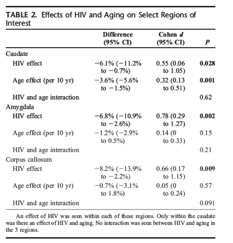
F2 and compared with HIV2 subjects (Fig. 2). Both increasing age and HIV infection were each associated with significant decreases in caudate volume, but no significant interaction was present. For every 10 years, HIV led to a 6% decrease in caudate volume, whereas aging resulted in a 4% decrease (Table 2). Overall, for a given age, caudate volumetric values
in a HIV+ subject were equivalent to a HIV- participant who was approximately 17 years older. For the amygdala and the corpus callosum, significant effects of HIV but not aging were observed. No significant age by HIV interactions were seen within these 2 regions (Table 2).
HAART Does Not Change Brain Volumetrics
Within the subset of the HIV+/HAART- that started medications, neuroimaging was performed both before and after starting HAART. The introduction of HAART led to a significant reduction in log10 plasma HIV VL (P <0.001) and an increase in CD4 cell count (P = 0.03; data not shown). However, there was no change in volumetric measures within select ROIs (ie, caudate) after starting HAART (see Figure, Supplemental Digital Content 3, http://links.lww.com/QAI/A265). Some improvements on neuropsychological performance testing were seen at the second assessment, but this difference was not statistically significant (P = 0.65). For each of the ROIs, there was no significant effect of self-reported duration of HAART on brain volume (data not shown).
Caudate Volume Atrophies After
Approximately 13 Years of HIV Infection
The difference in brain volume between the HIV+ and HIV- subjects suggests that following the time of self-reported infection, certain regions may be at increased risk for atrophy. Based on an exploratory model that uses thebroken-stick method, the estimated time (T) needed for the brain volume to transition from self-reported date of HIV+ infection to HIV-, the level for chronically HIV+ infection was 161 months (13.4 years), 95% confidence interval (3-300 months) (Fig. 3). Although a significant degree of variability was observed, a more gradual decline in brain volume fit the data better than a one implying abrupt atrophy (improvement in Akaike information criterion = 0.57).
| |
| |
| |
|
|
|