| |
Kidney Disease Review: The Top 10 Things Nephrologists Wish Every Primary Care Physician Knew
|
| |
| |
Download the PDF here
Mayo Clinic Proceedings February 2009
"Early recognition, evaluation, and appropriate treatment and/or referral are necessary to moderate the substantial morbidity and mortality that are often associated with diseases of the kidney."
Renal disease is commonly encountered by primary care physicians during their day-to-day visits with patients. Common renal disorders include hypertension, proteinuria, kidney stones, and chronic kidney disease. Despite their prevalence, many physicians may be unfamiliar with the diagnosis and initial treatment of these common renal disorders. Early recognition and intervention are important in slowing the progression of chronic kidney disease and preventing its complications. The evidence-based pearls in this article will help primary care physicians avoid common pitfalls in the recognition and treatment of such disorders and guide their decision to refer their patients to a specialist.
ACEI, angiotensin-converting enzyme inhibitor , ARB, angiotensin II receptor blocker , BUN, blood urea nitrogen , CKD, chronic kidney disease , GFR, glomerular filtration rate , LDL-C, low-density lipoprotein cholesterol , NSAID, nonsteroidal anti-inflammatory drug
Chronic kidney disease (CKD) is a widespread public health problem with substantial morbidity and mortality. Outcomes associated with CKD are progression to kidney failure, cardiovascular disease, and premature death. Learning to recognize CKD in its earliest stage and understanding what measures to undertake to prevent its progression and associated complications are important goals developed by the Kidney Disease: Improving Global Outcomes (KDIGO) initiative.1 The evaluation of difficult-to-treat hypertension and kidney stones are additional skills that primary care physicians will need to acquire. The evidence-based pearls in this article will help primary care physicians understand these concepts and avoid common pitfalls in the recognition and treatment of such disorders and guide their decision to refer their patients to a specialist.
1. A "Normal" Serum Creatinine Level May Not Be Normal. Normal creatinine values may vary among different laboratories, and some patients with serum creatinine levels within the "normal" range may have substantial reduction in kidney function. For example, a 66-year-old man with a serum creatinine level of 1.4 mg/dL (to convert to μmol/L, multiply by 88.4) would have an estimated glomerular filtration rate (GFR) of 54 mL/min per 1.73 m2. This perceived "normal" serum creatinine value actually corresponds to stage 3 CKD.1 Likewise, the rate of increase or trend of serum creatinine levels can also indicate CKD. For example, a 66-year-old man with a serum creatinine level of 0.8 mg/dL and an estimated GFR of 103 mL/min per 1.73 m2 would have an estimated GFR of less than 64 mL/min per 1.73 m2 should the serum creatinine level stabilize at 1.2 mg/dL. This would represent at least a 40% reduction in GFR. Estimates of GFR can only be based on steady-state serum creatinine values and cannot be used to accurately estimate GFR when it is changing. A rapid increase in the serum creatinine level from 0.8 to 1.2 mg/dL within 8 hours could reflect a GFR approaching zero in a patient with acute renal failure. The interpretation of serum creatinine level also depends on muscle mass, age, sex, height, and limb amputation. A patient with an amputation and a high-normal creatinine level may actually have advanced CKD. Commonly used GFR estimation equations, such as the Modification of Diet in Renal Disease calculator (www.mdrd.com), have not been validated in patients with normal GFR and in fact may underestimate actual GFR in healthy people.2 The take-home message is that recognition of CKD is important in order to identify and treat reversible causes of renal disease, slow the progression and avoid the complications of renal disease, and identify patients who may need dialysis or a renal transplant.1
2. Know the Medications That Spuriously Elevate the Serum Creatinine Level. In a number of scenarios, the serum creatinine level can increase without reflecting a change in the actual GFR. The antibiotic trimethoprim-sulfamethoxazole and the H2-blocker cimetidine are 2 commonly used drugs that decrease the secretion of creatinine. This can result in a self-limited and reversible increase in the serum creatinine level of as much as 0.4 to 0.5 mg/dL (depending on baseline serum creatinine level). Famotidine and ranitidine can likewise cause an increase but to a lesser degree. The antibiotic cefoxitin can spuriously increase the serum creatinine level by interfering with the colorimetric assay used to measure serum creatinine levels.3 In both instances, the blood urea nitrogen (BUN) typically does not change. As such, an increase in creatinine level suggests a true decrease in GFR only if accompanied by a corresponding increase in BUN levels.
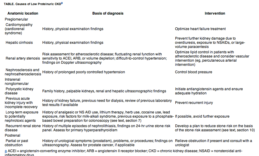
3. Patients With Decreased GFR or Proteinuria Should Be Evaluated to Determine the Cause; Positive Urine Dipstick Test Results for Protein Should Be Followed Up With a Spot Urine Protein to Urine Creatinine Ratio. Chronic kidney disease is defined as a kidney abnormality that persists for more than 3 months. Persons with a normal GFR may have CKD if they have persistent proteinuria or hematuria of renal origin. Despite the difficulty associated with accurate assessment of near-normal GFR, most would agree that a persistent GFR of less than 60 mL/min per 1.73 m2 would signify CKD. Proteinuria is initially detected by the urine dipstick test, which reflects the concentration of albumin in the urine. Because variations in urine flow and concentration can affect the semiquantitative dipstick determination, a quantitative estimation of proteinuria is required.The preferred method for quantitative estimation of protein excretion is the spot urine protein to creatinine ratio because it is accurate and more convenient than a 24-hour urine collection.4, 5, 6, 7 In patients at risk of proteinuria (eg, patients with diabetes), a spot urine albumin to creatinine ratio is helpful for detecting microalbuminuria, thereby guiding further therapy. Patients with diabetes who have microalbuminuria may be at higher risk of developing diabetic nephropathy. In a patient with a urine protein to creatinine ratio of 1 or more, the risk of progression of CKD is higher. In fact, any patient with an elevated ratio (≥1) should be evaluated for causes of glomerular disease, including diabetes, collagen vascular disease (eg, systemic lupus erythematosus), malignancy (eg, multiple myeloma), infections (eg, human immunodeficiency virus infection, syphilis, hepatitis B and C), and use of medications such as nonsteroidal anti-inflammatory drugs (NSAIDs).8 Solid tumors may also be associated with glomerular disease (membranous glomerulopathy), but such tumors are generally either already known or readily detectable by age-, risk-, and sex-appropriate cancer screening.9, 10 Patients with a high degree of proteinuria or active urinary sediment, especially in the setting of deterioration in kidney function, should be referred to a nephrologist for further evaluation and treatment. A lower degree of proteinuria in association with CKD (unmodified by antiangiotensin or antialdosterone agents) suggests a nonglomerular etiology (Table). This Table should not be considered to be all inclusive; instead, it provides a conceptual framework for approaching the identification of the numerous causes of nonglomerular CKD. Preglomerular, intrarenal, and postrenal causes may be associated with long-term reductions in renal function and lower degrees of proteinuria. The key to the diagnostic assessment of patients with these conditions includes, most importantly, a careful history and physical examination, followed by renal Doppler ultrasonography to detect renal artery stenosis, obstruction, and/or cysts.
4. In Patients With Early-Stage CKD, Periodic Evaluation and Intervention Are Appropriate to Slow the Progression of Renal Disease and Avoid Its Complications. In patients with CKD, it is imperative to slow the rate of disease progression. Nephrotoxic drugs, such as NSAIDs, aminoglycoside antibiotics, and radiocontrast agents, should be used with caution or avoided completely. Systemic blood pressure should be monitored frequently and controlled with a goal blood pressure of less than 130/80 mm Hg. A lower blood pressure may be desirable in patients with proteinuric CKD. A spot urine protein to creatinine ratio should be obtained periodically, with a ratio of 1 or more suggesting a higher risk of CKD progression.Angiotensin-converting enzyme inhibitors (ACEIs) and angiotensin II receptor blockers (ARBs) may slow the progression of CKD, especially in patients with proteinuria. In addition to lowering systemic blood pressure, ACEIs and ARBs also lower glomerular capillary blood pressure and protein filtration, which may contribute to their beneficial effect in slowing progression. They may also help reduce angiotensin II-mediated cell proliferation and fibrosis.11 Combination therapy with an ACEI and ARB may have a greater antiproteinuric effect than either drug alone, but patients should be monitored carefully for increases in serum creatinine and potassium levels.11, 12 Patients with progressive CKD are at risk of renal osteodystrophy. Phosphorus, calcium, and parathyroid hormone levels should be monitored closely in all patients with stage 3 to 4 CKD. Abnormalities in these values may indicate the need for dietary phosphate restriction, administration of oral phosphate binders, and/or the administration of vitamin D.Patients with CKD also have an increased risk of cardiovascular complications, including myocardial infarction.13 Aspirin (eg, 81 mg/d) and aggressive lipid-lowering strategies should be used, including 3-hydroxy-3-methylglutaryl (HMG) coenzyme A reductase inhibitors or statins, with a goal low-density lipoprotein cholesterol (LDL-C) level of 100 mg/dL or less (to convert to mmol/L, multiply by 0.0259). In patients with CKD and established coronary artery disease, some physicians recommend a goal LDL-C level of 70 mg/dL or less. Recently, several studies have suggested that statins may have the additional benefit of reducing proteinuria.4, 13 More studies are needed to determine whether tighter control of LDL-C levels translates into reduction of cardiovascular events or progression to end-stage renal disease.Early referral to a nephrologist should be considered if CKD progresses or if the patient has uncontrolled complications, including nephritic-range proteinuria, uncontrolled blood pressure, uncontrolled secondary hyperparathyroidism, or active urine sediment. Consultation and/or comanagement with a kidney disease care team is advisable for patients with stage 3 CKD (GFR, 30-59 mL/min/1.73 m2). All patients with a GFR of less than 30 mL/min per 1.73 m2 (stages 4-5) should be referred to a nephrologist.9
5. Do Not Automatically Discontinue an ACEI or ARB Solely Because of a Small Increase in the Serum Creatinine or Potassium Level. Two important drugs in the treatment of patients with CKD are ACEIs and ARBs. In management of proteinuric kidney diseases, ACEIs or ARBs are used not only to optimize blood pressure control (Joint National Commission VII) but can be titrated up in patients with proteinuria. They are also the drugs of choice to prevent progression of proteinuric CKD.12, 14, 15 Patients who begin taking or have a dose increase in ACEIs or ARBs may experience an increase in the serum creatinine level. Although an increase of 20% to 30% is acceptable,16 it is important to confirm that the serum creatinine level stabilizes at the higher value and does not continue to increase. Also, an increase in serum potassium levels is sometimes seen in patients who are taking ACEIs or ARBs. A serum potassium level of up to 5.5 mEq/L (to convert to mmol/L, multiply by 1.0) is acceptable as long as it is stable and as long as the patient is aware of the need for dietary potassium restriction and will not be exposed to additional medications, such as spironolactone, that may exacerbate hyperkalemia. Follow-up serum creatinine and potassium levels should be ordered within 1 week. In patients with an increase in creatinine level of more than 20% to 30% or in those with uncontrollable hyperkalemia, the ACEI or ARB should be discontinued or titrated to a lower dose. Frequent monitoring is necessary. Furthermore, diuretics that work with reduced GFR, such as furosemide or metolazone, are useful agents in the treatment of hypertension and hyperkalemia in CKD. However, it is important to note that volume depletion may make a patient more susceptible to an ACEI- or ARB-induced increase in the serum creatinine level.
6. Anemia in Patients With CKD Should Be Treated With Erythrocyte-Stimulating Agents Such as Recombinant Human Erythropoietin But Should Not Be Overtreated. In patients with CKD, anemia has been associated with fatigue, reduced exercise tolerance, dyspnea, left ventricular hypertrophy, left ventricular systolic dysfunction, and an increased risk of cardiovascular events (stroke and myocardial infarction).17, 18 As such, the National Kidney Foundation Dialysis Outcomes Quality Initiative (K/DOQI) guideline for the treatment of anemia of CKD recommends that the hemoglobin target should be between 11 and 12 g/dL (to convert to g/L, multiply by 10) and should not exceed 13 g/dL.18 Erythropoietin should be discontinued in patients with a hemoglobin level of 13 g/dL. In a number of well-publicized articles, patients taking erythropoietin who were corrected to normal or near-normal hemoglobin levels were at higher risk of cerebrovascular events, thrombosis, and hypertension.18, 19, 20, 21 All patients with anemia should be evaluated for other reversible causes, including vitamin deficiencies and iron deficiency. Routine evaluation would include a reticulocyte count and measurement of serum vitamin B12 and folate, serum iron, ferritin, and total iron-binding capacity. Patients (especially males and nonmenstruating females) with iron deficiency should undergo a gastrointestinal evaluation with endoscopy and colonoscopy. Iron deficiency must be corrected if erythrocyte-stimulating agents are to be effective.
7. Phosphate-Containing Bowel Preparations Should Be Used With Caution. Although sodium phosphate bowel preparation agents are effective and more convenient than other agents on the market, several recent studies have suggested that they can cause acute phosphate nephropathy, leading to acute renal failure or worsening of CKD.22, 23, 24 A 2005 study reported 21 cases of acute phosphate nephropathy in patients who had undergone a recent colonoscopy and who had taken a sodium phosphate bowel preparation.25 Although the pathophysiology was not fully understood, it was thought to be secondary to substantial fluid shifts and electrolyte changes. On the basis of their case evaluation, the authors of this study speculated that potential etiologic factors included inadequate hydration, increased patient age, a history of hypertension, and current use of an ACEI or ARB. Subsequently, several professional societies have developed guidelines for the use of sodium phosphate bowel preparations.26, 27 In addition to the potential etiologic factors identified in the 2005 study,25 patients with CKD or chronic heart failure and those taking NSAIDs or diuretics would also appear to be at higher risk of acute phosphate nephropathy. For these patients, the guidelines recommend an alternative bowel preparation agent, polyethylene glycol, which is not associated with volume shifts and electrolyte abnormalities. Because patients must drink a large volume of this alternative bowel preparation agent, adherence can be an issue. In deciding whether to use a bowel preparation agent and in selecting which one to use, clinicians should carefully weigh the benefits and risks for each individual patient.
8. Patients With Severe CKD Should Avoid Magnesium- or Aluminum-Containing Oral Preparations. Concomitant Use of Citrate-Containing Preparations and Aluminum-Containing Oral Preparations Is Potentially Hazardous Because It Can Lead to Acute Aluminum Toxicity. In patients with severe CKD, over-the-counter antacids that contain aluminum and magnesium (eg, Maalox and Mylanta) should be avoided. Other medications, such as the cathartic agent magnesium citrate, should likewise be used with caution. Indiscriminate use of magnesium-containing preparations can lead to severe hypermagnesemia. In addition, decreased renal function also increases the risk of aluminum accumulation and subsequent bone disease and neurotoxicity.28, 29 Chronic aluminum toxicity has been linked to sporadic Alzheimer disease and other neurodegenerative disorders30; however, this link is highly controversial. Moreover, certain medications, such as calcium citrate, potassium citrate, and sodium citrate, markedly enhance aluminum absorption from the gut. A number of studies have suggested that the concomitant use of citrate-containing preparations and aluminum hydroxide is potentially hazardous.31, 32, 33 In one such study, Kirschbaum and Schoolwerth34 reported the development of a rapidly progressive encephalopathy marked by confusion, myoclonus, seizures, coma, and death in a group of women with renal failure who received an oral solution of citrate and aluminum hydroxide gel concurrently. Given these findings, patients with CKD who need to take a medication such as sodium citrate to prevent kidney stones or another citrate-containing preparation should not take aluminum hydroxide-containing medications. Because of the universal availability of such over-the-counter antacids, these patients should be advised of this specific contraindication.
9. Although Most Patients With Hypertension Should Not Be Screened for Secondary Hypertension, Certain Clinical Clues May Suggest the Presence of an Underlying Cause That, When Addressed, May Resolve or Improve the Patient's Hypertension. From an epidemiologic standpoint, secondary hypertension is relatively uncommon. Of patients who are diagnosed as having hypertension in a primary care clinic, 95% have primary or essential hypertension, and only 5% have a secondary cause.35 In a study at a hypertension clinic, secondary hypertension accounted for only 9% of all patients seen.36 Severe or difficult-to-control hypertension, hypertension that suddenly develops or suddenly worsens, or hypertension that is associated with other clinical findings may indicate secondary hypertension.37, 38 Hypokalemia may suggest primary aldosteronism; however, in 1 series, hypokalemia was present in only 25% of patients diagnosed as having primary hyperaldosteronism.36 Headaches, palpitations, and sweats may suggest pheochromocytoma. Moon facies and/or striae may suggest Cushing syndrome, and a history of snoring in an obese patient may suggest obstructive sleep apnea. A bruit on one side of the abdomen may indicate renal artery stenosis. Worsening blood pressure or renal function on initiation of an ACEI or ARB may also suggest renal artery stenosis. Patients with peripheral vascular, cardiovascular, or cerebrovascular disease are at risk of also having renovascular disease. Other potentially correctable systemic conditions that can cause hypertension include hypothyroidism and hyperparathyroidism.Each of these findings or the already described historical features may suggest a secondary cause of the hypertension. Treatment of the underlying disorder can improve the patient's blood pressure and, in some instances, may resolve the patient's hypertension. Clinicians should determine whether the patient's medications (eg, NSAIDs, birth control pills, or certain decongestants) or lack of adherence to dietary restrictions (eg, ingestion of sodium and sodium-containing foods) is contributing to his or her suboptimal control and should review the patient's antihypertensive regimen for appropriateness. Assessment for target organ damage is also appropriate to determine cardiovascular risk. Determining when to evaluate for a secondary cause of hypertension is based on the pretest probability or the likelihood that 1 or more of the above historical features or physical findings are present. A stepwise approach should be used.
10. In Patients With Recurrent Stone Disease, an Indepth Metabolic Evaluation Is Needed to Identify and Treat Modifiable Risk Factors, Thereby Preventing Further Episodes and/or Promoting Stone Dissolution. Nephrolithiasis, a common problem encountered by primary care physicians, can cause substantial morbidity. The likelihood that stone disease will develop in a man by age 70 years is as high as 1 in 8.39 The 10-year recurrence rates after a first calcium oxalate stone can be as high as 50% without treatment. It can be much higher in patients with a metabolic risk. As such, patients with an initial stone should undergo risk assessment. Patients with a family history of stones, concomitant gastrointestinal disease such as inflammatory bowel disease, frequent urinary tract infections, or a history of nephrocalcinosis should be referred to a nephrologist for further evaluation. Patients without these risk factors should have a simplified evaluation that includes a dietary history, a review of medications that can promote stone development, urinalysis, and measurement of levels of serum calcium, phosphorous, electrolytes, and uric acid.40 Compositional stone analysis is an integral part of the metabolic evaluation because it can provide guidance for therapy.41 The first-time diagnosis of a single calcium-containing kidney stone in a patient without a family history of kidney stones and with normal findings on an initial metabolic evaluation can be managed by increasing fluid intake to maintain a urine output of about 2 L/d. If a uric acid or cystine stone is detected on stone analysis, further metabolic evaluation with a 24-hour urine stone risk profile (eg, UroRisk or StoneRisk Panel [Mission Pharmacal Company, San Antonio, TX]) is warranted to guide both dietary and medical interventions.42 Likewise, in patients without a stone available for analysis or in patients with multiple calcium stones, further metabolic evaluation, including the 24-hour urine stone risk profile, is recommended.
One Additional Point: Cyclosporine and Tacrolimus, Drugs Commonly Used in Patients With Renal Allografts, Have Many Drug-Drug Interactions
Every new medication (prescribed, over-the-counter, and herbal) that is given to a patient with a renal allograft should be reviewed to determine if it will interact with his or her transplant medications (eg, tacrolimus or cyclosporine). Some medications may decrease calcineurin inhibitor levels; others may cause cyclosporine or tacrolimus toxicity.43 In both instances, the health of the patient or the success of the renal allograft may be jeopardized. For example, St Johns Wort, a herbal preparation, may decrease cyclosporine levels substantially, potentially resulting in acute rejection. Several other commonly prescribed medications that can be associated with reduced cyclosporine levels include rifampin, phenytoin, and carbamazepine. In contrast, diltiazem, verapamil, and erythromycin may increase cyclosporine levels. Cyclosporine can interfere with the metabolism of certain statins such as simvastatin, increasing the risk of statin-induced rhabdomyolysis. If any medication must be given to a patient with a renal allograft who is taking tacrolimus or cyclosporine, a careful review of its interaction with these medications should be made. If a drug must be used that has a considerable interaction, dose adjustment may be needed with careful follow-up of the calcineurin inhibitor levels. Careful monitoring is the rule.
Conclusion
Renal disease is commonly encountered by primary care physicians. Early recognition, evaluation, and appropriate treatment and/or referral are necessary to moderate the substantial morbidity and mortality that are often associated with diseases of the kidney.
Questions About Common Renal Problems
1. A 65-year-old man with stage 3 chronic kidney disease (CKD) is noted to have proteinuria on dipstick testing and a subsequent urine protein to creatinine ratio of 0.7. The patient's blood pressure has been in the range of 130/80 mm Hg while he has been taking a moderate dose of an angiotensin-converting enzyme inhibitor (ACEI). You increase his ACEI dose and see him in follow-up a week later. Although his blood pressure has improved and his urine protein to creatinine ratio has decreased to 0.5, you note that his serum creatinine level has increased from 1.4 to 1.7 mg/dL. Which one of the following is the next best step in the treatment of this patient?
a.Stop the ACEI and initiate dihydropyridine calcium channel blocker therapy
b.Lower the ACEI dose back to baseline and initiate dihydropyridine calcium channel blocker therapy
c.Continue the current regimen but recheck the patient's serum creatinine level in 1 week
d.Add an angiotensin II receptor blocker (ARB) to his current regimen
e.Stop the ACEI and start an ARB
2. A 55-year-old man presents with a blood pressure of 160/90 mm Hg and a 15-year history of hypertension and long-standing hypercholesterolemia treated with a medication regimen of 50 mg/d of losartan, 10 mg/d of amlodipine, 100 mg/d of atenolol, and 25 mg/d of hydrochlorothiazide. He is otherwise feeling well and denies headaches or flushing. During the past year, his blood pressure has been more difficult to control (averaging 160/90 mm Hg), and, 1 week previously, the dosage of losartan was increased to 100 mg/d. His serum electrolyte levels are normal, and his serum creatinine level is 1.1 mg/dL. Which one of the following should be the next step in the management of this patient?
a.Initiate 12.5 mg/d of spironolactone
b.Obtain an aldosterone-renin ratio; refer for ultrasonography to rule out renal artery stenosis
c.Increase the dosage of hydrochlorothiazide to 50 mg/d
d.Initiate 5 mg of minoxidil twice daily
e.Perform magnetic resonance imaging of the abdomen
3. Which one of the following statements is false about the management of a patient with a serum creatinine level of 2.2 mg/dL?
a.Certain over-the-counter medications, such as nonsteroidal anti-inflammatory drugs (NSAIDs) and antacids, should be avoided
b.Both ACEIs and ARBs can be used in patients with proteinuria
c.Serum phosphorus, calcium, and parathyroid hormone levels should be monitored closely
d.Treating anemia with recombinant human erythropoietin to the normal hemoglobin range has been shown to reduce morbidity and mortality
e.In addition to lowering cholesterol, statins may also reduce proteinuria
4. Which of the following patients is likely to have CKD?
a.A 55-year-old woman with a serum creatinine level of 1.2 mg/dL
b.A 45-year-old man with bilateral below-the-knee amputations and a serum creatinine level of 1.1 mg/dL
c.An 80-year-old woman with a serum creatinine level of 1.0 mg/dL
d.A 70-year-old man with some muscle wasting who weighs 65 kg and has a serum creatinine level of 1.1 mg/dL
e.All of the above
5. Which one of the following statements is false about the management of a patient with a kidney stone?
a.Compositional stone analysis identifying the type of stone will help guide therapy
b.A first calcium stone can be simply managed by increasing fluid intake to maintain a urine output of 2 L/d
c.If a stone is not recovered and sent for analysis, a 24-hour urine stone risk profile should be ordered
d.A first uric acid or cystine stone can be simply managed by increasing fluid intake to maintain a urine output of 2 L/d
e.Patients with inflammatory bowel disease are at increased risk of kidney stones
| |
| |
| |
|
|
|