| |
Inflammation, Activation Associated with Reduced Functional Capacity in HIV+
|
| |
| |
Download the PDF here
"we detected an association between high IL-6 level and low functional status and a trend toward higher hs-CRP and TNF-α levels suggesting an overall inflammatory milieu among functionally impaired persons.....our findings suggest that a lower nadir CD4+ T-cell count prior to initiation of antiretroviral therapy and continued perturbations in CD4+ T-cell proportions during antiretroviral therapy place HIV-infected patients at risk for functional impairment through chronic immune activation and inflammation."
from Jules: exercise & diet are a key component to addressing inflammation & functioning
Functional Assessments in study: "A frailty phenotype was assessed.....Short Physical Performance Battery assessed tandem stand, walking speed, and sit-stand test time.....Four hundred meter walk time was measured"
IAS/Rome/2011: Frailty Rate in HIV+ mid-50 Women Matches Rate in 70-Year-Olds Without HIV - written by Mark Mascolini - (07/20/11)
"Our study was the first comparison of markers of immune activation, immunosenescence, and microbial translocation in HIV-infected persons with phenotypes defined by functional capacity. We identified a distinct clinical phenotype on the basis of physical function and frailty assessment that is associated with markers of inflammation and immune activation that predict poor prognosis in HIV-infected persons. A measurement of physical function can be easy, fast, and inexpensive. Future interventions targeted at decreasing immune activation and inflammation in HIV-infected persons should assess the clinical impact of therapy through measures of physical function."
"In HIV-uninfected persons, chronic inflammation, antigen stimulation, and age lead to immune remodeling manifested by a decreased CD4+ T-lymphocyte level, an increased CD8+ T-lymphocyte level, and expansion of CD28- T-lymphocytes with shortened telomeres [3]. These changes have been associated with frailty and disability in HIV-uninfected elderly individuals"
"........Functional status incorporates the concomitant processes of aging, inflammation, HIV infection, comorbid diseases, and lifestyle. Using a case-control design, we demonstrated significant independent associations of functional impairment with IL-6 and CD8+ T-cell activation in patients receiving effective combination antiretroviral therapy. Our findings add to a growing body of evidence for the association of chronic immune activation and inflammation with the development of non-AIDS morbidity and mortality [30-32]. To our knowledge, this is the first evidence linking physical function and frailty to immune activation and inflammation among persons with HIV infection."
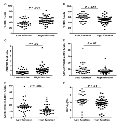
"Markers of inflammation, T-cell activation, microbial translocation, immunosenescence, and immune recovery were used to estimate functional status.......HIV-infected persons who were aged 45-65 years, had a plasma HIV-1 RNA load of <48 copies/mL, and were receiving antiretroviral therapy......359 HIV+ completed functional testing......Thirty-three (9%) were identified as having low functional status, 186 (52%) were identifies as having moderate functional status, and 140 (39%) were identified as having high functional status......Low-function cases had a lower nadir CD4+ T-cell count, a greater number of comorbid conditions.....Although total CD4+ T-cell count was not significantly different between low-functioning and high-functioning groups (Table 1), a lower CD4+ T-cell percentage, a higher CD8+ T-cell percentage, and a lower ratio of CD4+ to CD8+ T cells were associated with a greater odds of low-functioning status. Significant differences were detected between low- and high-functioning groups with respect to CD4+ T-cell percentage (Figure 1A), CD8+ T-cell percentage (Figure 1B), and ratio of CD4+ to CD8+ T cells (Figure 1C)."
"Similar to prior studies of frailty and disability among HIV-uninfected elderly individuals, we detected an association between high IL-6 level and low functional status and a trend toward higher hs-CRP and TNF-α levels, suggesting an overall inflammatory milieu among functionally impaired persons. Moreover, increased CD8+ T-cell activation was also associated with functional impairment among HIV-infected persons. Higher IL-6 level is closely linked to frailty, disability, and mortality among HIV-uninfected elderly individuals [3, 7], while CD8+ T-cell activation is associated with disease progression and poor immune recovery in HIV-infected persons [17]. However, the role of CD8+ T-cell activation in physical function impairment is largely unexplored in HIV-infected and HIV-uninfected populations [8]."
Figure 1. Comparison of T-lymphocyte subsets and immune activation (CD38 and HLA-DR expression on CD4+ and CD8+ T cells and soluble CD14 [sCD14] level) between low- and high-functioning groups. Means and standard errors are represented by bars and error bars, respectively. P values were determined by unadjusted comparison of means in a mixed-effects model to account for clustering from matched design.
---------------------------------------------
Association of Functional Impairment with Inflammation and Immune Activation in HIV Type 1-Infected Adults Receiving Effective Antiretroviral Therapy
Journal of Infectious Diseases July 2013
Kristine M. Erlandson,1,2 Amanda A. Allshouse,3 Catherine M. Jankowski,2 Eric J. Lee,1 Kevin M. Rufner,4
Brent E. Palmer,5 Cara C. Wilson,1 Samantha MaWhinney,3 Wendy M. Kohrt,2 and Thomas B. Campbell1
1Division of Infectious Diseases, 2Division of Geriatric Medicine, 3Department of Biostatistics and Informatics, University of Colorado, Denver, 4Division
of Gastroenterology and Hepatology and 5Division of Allergy and Clinical Immunology, Department of Medicine,
Presented in part: 6th International AIDS Society Conference on HIV Pathogenesis,
Treatment and Prevention, Poster TULBPE029 Rome, Italy, July 2011
Abstract
Background. The relationships of inflammation, immune activation, and immunosenescence markers with functional impairment in aging human immunodeficiency virus type 1 (HIV-1)-infected persons are unknown.
Methods. HIV-infected persons who were aged 45-65 years, had a plasma HIV-1 RNA load of <48 copies/mL, and were receiving antiretroviral therapy underwent standardized functional testing. In a nested case-control analysis, low-functioning cases were matched (1:1-2) by age, sex, and HIV-1 diagnosis date to high-functioning controls. Markers of inflammation, T-cell activation, microbial translocation, immunosenescence, and immune recovery were used to estimate functional status in conditional logistic regression. Primary analyses were adjusted for CD4+ T-cell count, smoking, and hepatitis.
Results. Thirty-one low-functioning cases were compared to 49 high-functioning controls. After statistical adjustment, lower proportions of CD4+ T cells and higher proportion of CD8+ T cells, higher CD38/HLA-DR expression on CD8+ T cells, and higher interleukin-6 were associated with a significantly greater odds of low functional status (odds ratio, ≥ 1.1 for all analyses; P ≤ .03). Other inflammatory, senescence, and microbial translocation markers were not significantly different (P ≥ .11 for all analyses) between low-functioning and high-functioning groups.
Conclusions. Functional impairment during successful antiretroviral therapy was associated with higher CD8+ T-cell activation and interleukin 6 levels. Interventions to decrease immune activation and inflammation should be evaluated for their effects on physical function and frailty.
A cornerstone of successful aging is the ability to maintain functional independence [1, 2]. A key and often modifiable element of functional independence is the preservation of physical function. Physical function impairments in older adults increase the risk of morbidity and mortality. Among aging individuals without human immunodeficiency virus type 1 (HIV-1) infection, declines in physical function and development of frailty are strongly associated with inflammation and immune system dysfunction, including immune activation, immunosenescence, and altered levels of T-lymphocyte subsets [3-8].
Aging with HIV infection is associated with early decline in physical function and development of frailty [9]. Early development of frailty or a frailty-like syndrome has been demonstrated in several HIV cohorts, although findings have been most apparent in those who are not receiving antiretroviral therapy or have the lowest CD4+ T-cell counts [10-14]. Performance-based measures targeted at the upper end of the functional spectrum among middle-aged and older adults with HIV infection have identified greater than expected impairments in walking speed, balance, ability to rise from a chair, and peak exercise capacity [15, 16].
Given that immune activation and inflammation have been associated with impaired physical function and frailty in older non-HIV-infected persons and because HIV infection is associated with chronic immune activation and inflammation despite otherwise successful antiretroviral therapy [17], we hypothesized that functional impairment during HIV infection is associated with markers of immune activation and inflammation. Because immunosenescence is associated with frailty in non-HIV-infected elderly patients and because immunosenescence and microbial translocation have been associated with chronic HIV infection, we further hypothesized that markers of immunosenescence and microbial translocation would also be associated with functional impairment.
METHODS
Study Population
All individuals receiving care for HIV infection in the Infectious Diseases Group Practice at the University of Colorado were evaluated for participation. Individuals were eligible if they were aged 45-65 years, able to consent and participate in study procedures, and taking a minimum of 2 antiretroviral drugs for at least 6 months with 1 undetectable plasma HIV-1 RNA load measurement (lower limit of detection, <48 copies/mL) and no plasma HIV-1 RNA load of >200 copies/mL in the prior 6 months. Approval was obtained by the Colorado Multiple Institutional Review Board, and informed consent was obtained from all participants.
Clinical Assessments
All participants completed a standardized interview, and available medical records were reviewed. Self-reported time since HIV diagnosis, nadir CD4+ lymphocyte count, and HIV treatment history were confirmed by medical records when available. The presence or absence of the following comorbidities were determined by medical records: seizure disorder, dementia, stroke, neuropathy, psychiatric disease, arthritis, osteopenia or osteoporosis (prior stress fracture or T score of less than -1 on bone densitometry scan), diabetes, kidney disease (creatinine clearance rate of <30 mL/min, by the Cockcroft-Gault formula), malignancy (excluding nonmelanoma skin cancer), solid organ transplant, lung disease, hypertension, cardiovascular disease, viral hepatitis (presence of hepatitis B virus surface antibody or hepatitis C virus antibody), and chronic liver disease. Medications were determined by medical record review and self-report. Laboratory values were the most recent values available in the medical record. The Veterans Aging Cohort Study Index was calculated using the following parameters as previously described: CD4+ T-cell count, viral load, age, aspartate aminotransferase level, alanine aminotransferase level, platelet level, hemoglobin level, hepatitis C, and estimated glomerular filtration rate [18, 19]. Of a possible 164 points, higher values indicate greater mortality risk, and scores of ≤34 are associated with the lowest mortality [18, 19].
Functional Assessments
A frailty phenotype was assessed as previously described by Fried et al [20]. Shrinking was defined as unintentional weight loss of ≥10 pounds or a decrease of 5% of body weight in the last year (data were self-reported and verified by records when available). Exhaustion was defined as feeling 4-5 times per week that "everything I do is an effort" or that "sometimes I just cannot get going" [11, 12, 14, 20]. Low activity was defined as being "limited a lot" in vigorous physical activities and was measured by self-report using the Short Form 36 health survey [11, 12, 14]. Weakness was assessed as the average of 3 dominant hand grip measurements obtained by a single Lafayette dynamometer, applying previously defined sex and body mass index cutoffs [20]. Slowness was measured on the basis of 4.5-m walk time, with men ≤173 cm and women ≤159 cm in height who took ≥7 seconds and men >173 cm and women >159 cm in height who took ≥6 seconds considered to be slow [20]. One point was given for each abnormality. A total of 3-5 points was considered indicative of low functional status; 1 or 2 points, moderate functional status; and 0 points, high functional status [20].
The Short Physical Performance Battery assessed tandem stand, walking speed, and sit-stand test time, with 0 points indicating inability to complete a task and 4 points indicating performance within the expected range [21]. Tandem stand was measured by the ability to stand heel to toe for 10 seconds, walking speed was assessed by the faster of two 4-m walks at usual pace, and sit-stand test time was assessed by 5 repetitions of going from a seated position to a standing position without use of the arms [21]. A score of <9 is highly predictive for subsequent disability and was considered indicative of low functional status; 9-11 points, moderate functional status; and 12 points (no deficits), high functional status [21, 22].
Four hundred meter walk time was measured on a set walking course by asking the participant to walk as quickly as possible to complete the distance [23, 24].
We defined high function as the ability to complete a 400-m walk and no deficits on either the frailty phenotype or the Short Physical Performance Battery. Low function was defined as a score of ≥3 on the frailty phenotype or a score of <9 on the Short Physical Performance Battery with at least 1 deficit on the opposing test. Moderate function was defined as at least 1 deficit on the frailty phenotype or Short Physical Performance Battery without meeting the definition of low function.
Flow Cytometric Assays
For measurement of HLA-DR and CD38 expression, whole blood was stained with fluorescently labeled anti-CD3/CD4/CD38/HLA-DR or anti-CD3/CD8/CD38/HLA-DR monoclonal antibodies (BD Biosciences, San Jose, CA). Cells were incubated for 30 minutes at room temperature, red blood cells (RBCs) were lysed, and remaining cells were fixed by the addition of 450 μL of FACS Lysing solution (BD Biosciences) before they were analyzed.
For CD28 expression, RBCs were lysed before staining by adding 4 mL of PharmLyse (BD Biosciences, San Jose, CA) to 1 mL of whole blood. The blood was incubated for 5 minutes at room temperature and centrifuged at 300 x g for 5 minutes to pellet the white blood cells. The cells were washed with FACS buffer (2% human serum in phosphate-buffered saline), and the pellet was resuspended in 100 μL of FACS buffer. The staining cocktail containing fluorescently labeled anti-CD3, -CD4, -CD8, and -CD28 monoclonal antibodies was added, and the cells were incubated at 4°C for 20 minutes. Cells were washed once with FACS buffer, resuspended in 100 μL of fixation buffer (1% paraformaldehyde), and stored at 4°C until analysis. A fluorescence minus 1 control that did not contain CD28 was included.
All flow cytometric assays used a FACSCalibur flow cytometer (BD Immunocytometry Systems, San Jose, CA). The senescence panel was acquired using Cell Quest Pro software (BD Immunocytometry Systems, San Jose, CA), and 100 000 events were collected. The percentage of CD28 expression on CD4+ and CD8+ T cells was determined using the fluorescence minus 1-stained cells to set the positive gate. CD38 and HLA-DR expression was analyzed using settings (voltage and compensation) determined by FACSComp, using CaliBRITE beads (BD Biosciences, San Jose, CA). Samples were analyzed using the HLA-DR/CD38 Multiset Algorithm, which automatically gates and determines the percentage of HLA-DR+/CD38+ CD4+ and CD8+ T cells.
Quantitative Polymerase Chain Reaction (PCR) Assays
Leukocyte telomere length was measured in cryopreserved whole blood. Genomic leukocyte DNA was isolated using Perfect Pure DNA Blood kit from 5 PRIME (Fischer Scientific, Pittsburgh, PA). Quantitative real-time amplification of the telomere sequence was performed by Cawthon's method [25], with modifications as described by O'Callaghan et al [26] to obtain absolute telomere length. Each sample was analyzed in quadruplet, and the interclass correlation was 0.89 (95% confidence interval [CI], .86-.91).
We isolated 16S ribosomal DNA (rDNA) from cryopreserved
ethylenediaminetetraacetic acid plasma, using a modified DNeasy extraction kit (Qiagen, Valencia, CA). The 16S rDNA was lysophilized and then quantified using real-time PCR by methods previously described elsewhere [27]. Samples were analyzed in duplicate; the coefficient of variation was 18.3%. Variance of pooled controls had a mean cycle threshold of 41.2 (95% CI, 39.4-43.0); 39.4 was set as the 0 cutoff, and all values were normalized to 39.4 cycles.
Measurement of Soluble Molecules in Plasma and Serum
Commercially available enzyme-linked immunosorbent assays were used to measure serum interleukin 6 (IL-6) and tumor necrosis factor α (TNF-α; R&D Systems, Minneapolis, MN), plasma soluble CD14 (sCD14; R&D Systems, Minneapolis, MN), lipopolysaccharide (LPS)-binding protein (LBP; Hycult, Plymouth Meeting, PA), endotoxin core IgM antibodies (endoCAb; Hycult, Plymouth Meeting, PA), and intestinal fatty acid-binding protein (i-FABP; R&D Systems, Minneapolis, MN). Highly sensitive C-reactive protein (hs-CRP) was measured by immunoturbidimetrics (Beckman Coulter, Brea, CA). Plasma LPS concentration was evaluated in triplicate and measured with the Endotoxin Detection Kit (Lonza, Walkersville, MD) after dilution of plasma at a ratio of 1:10 in endotoxin-free water (Hyclone, Logan, UT) and incubation at 80°C for 15 minutes. All assays followed the instructions specified by the manufacturer. The coefficient of variation was 1.2% for sCD14 and 17.6% for LPS.
Study Design and Data Analysis
A nested case-control study included all persons identified as having a low functional status as cases. Each case was matched to a high-functioning control. If >1 high-functioning control was identified, cases were matched to 2 controls until the target sample size of 80 subjects was reached. Matching was by rank order of age within 2 years, sex, and time since HIV diagnosis (prior to 1996 or after 1996). Thirteen cases were matched to 1 control and 18 cases were matched to 2 controls. All persons included as cases and controls were asked to return for a second visit for collection of blood samples. If participants had an infection or other acute illness, blood samples were not collected until a minimum of 14 days after symptom resolution. Persons who had taken oral corticosteroids within the prior 4 weeks were excluded from the case-control analysis. Blood samples were not collected from individuals who did not participate in the case-control analysis.
Odds ratios (ORs) and 95% CIs are presented from conditional logistic regression for the primary analysis, in which the conditional odds of low functional status was estimated for each measure, with adjustment for the most recent CD4+ T-cell count, viral hepatitis (positive hepatitis B virus surface antibody and/or hepatitis C virus antibody), and smoking. Mean differences and 95% CIs from mixed model comparisons are reported for secondary analyses, where continuous measures are summarized as mean values and 95% CIs from mixed model with matching identifier treated as a cluster and adjustment for most recent CD4+ T-cell count, viral hepatitis, and smoking. Skewed data (IL-6, hs-CRP, and 16S rDNA levels) were log transformed prior to analysis. Categorical data are presented as frequencies and percentages. The monotonic relationship between CD4+ and CD8+ T-cell activation and CD4+ T-cell count was assessed with the Spearman correlation coefficient. Analyses were performed in SAS v9.2 (Cary, NC). Data were collected and managed with Research Electronic Data Capture (REDCap), hosted at the University of Colorado [28].
RESULTS
Study Population
Between February and November 2010, 542 HIV-infected persons met eligibility requirements and were asked to participate: 171 either did not respond to correspondence or were not interested, 2 died prior to obtaining consent, 369 consented, and 359 completed functional testing. Thirty-three (9%) were identified as having low functional status, 186 (52%) were identifies as having moderate functional status, and 140 (39%) were identified as having high functional status. One low-functioning person moved from the area, and the other could not be contacted. A total of 31 low-functioning cases were matched to 49 high-functioning controls and underwent laboratory evaluations at the second study visit. Overall, participants were 85% male and 74% white, with a median age of 50.8 years (interquartile range, 47.7-55.7 years), and a median CD4+ lymphocyte count of 551 cells/μL (interquartile range, 361-768 cells/μL). The HIV-1 RNA load in 95% of subjects was below the limit of detection [29].
The low- and high-functioning groups were similar with respect to baseline demographic characteristics, with the exception that low-functioning cases were more likely to be current smokers (Table 1). Low-function cases had a lower nadir CD4+ T-cell count, a greater number of comorbid conditions, higher Veterans Aging Cohort Study Index scores, and were prescribed a greater number of nonantiretroviral medications (Table 1). Three subjects had plasma HIV-1 RNA detected by routine laboratory monitoring after the screening visit (1 low-functioning case had 491 copies/mL, and 2 high-functioning controls had 78 and 1670 copies/mL). One low-functioning case had a prior liver transplant and was receiving cyclosporine and sirolimus at the time of evaluation. Although total CD4+ T-cell count was not significantly different between low-functioning and high-functioning groups (Table 1), a lower CD4+ T-cell percentage, a higher CD8+ T-cell percentage, and a lower ratio of CD4+ to CD8+ T cells were associated with a greater odds of low-functioning status (Table 2). Significant differences were detected between low- and high-functioning groups with respect to CD4+ T-cell percentage (Figure 1A), CD8+ T-cell percentage (Figure 1B), and ratio of CD4+ to CD8+ T cells (Figure 1C).
Markers of Immune Activation and Inflammation
To determine the variability in CD38/HLA-DR expression on CD8+ T cells, 20 subjects had a repeat blood draw 2-5 days after the initial blood draw. Among these 20 subjects, interclass correlation was 0.88 (95% CI, .73-.95).
CD8+ but not CD4+ T-cell expression of CD38/HLA-DR was associated with a greater odds of having low functional status and remained significant after adjustment for CD4+ T-cell count, smoking, and hepatitis B or C (Table 2). Low-function persons had higher percentages of CD8+ and CD4+ T cells coexpressing CD38 and HLA-DR as compared to high-functioning persons (Figure 1D and 1E). In multivariate analyses with adjustment for CD4+ T-cell count, smoking, and hepatitis, CD38/HLA-DR on CD8+ T cells remained significantly higher among low-functioning persons (adjusted difference, 4.8; 95% CI, 1.4-8.3; adjusted P = .02), but significant differences in CD4+ T-cell activation were no longer detected (adjusted difference, 2.7; 95% CI, -.09 to 5.4; adjusted P = .07). Among the 3 subjects with detectable plasma HIV-1 RNA (Table 1), CD8+ T-cell expression of CD38/HLA-DR increased with increasing plasma HIV-1 RNA concentration (7% in a control with 78 copies/mL, 17% in a case with 491 copies/mL, and 25% in a control with 1670 copies/mL). CD8+ T-cell expression of CD38/HLA-DR remained higher in low-functioning cases (17.6% ± 1.4%) than in high-functioning controls (12.6% ± 1.2%) when the 3 subjects with detectable plasma HIV-1 RNA were excluded (P = .003). Among all subjects, higher CD4+ T-cell activation (r = -0.41, P < .001) and higher CD8+ T-cell activation (r = -0.38, P = .002) were moderately inversely correlated with CD4+ T-cell count.
Higher plasma levels of sCD14 were associated with a greater odds of low functional status, but differences were not robust after adjustment (Table 2). Similarly, plasma sCD14 concentration was higher among low-functioning as compared to high-functioning persons in univariate analysis (Figure 1F) but was not significant after multivariate analysis (adjusted difference, 0.3; 95% CI, .5-6.0; adjusted P = .09).
Although each log increase in IL-6 level increased the odds of low functional status by 1.2 (95% CI, 1.02-1.51; P = .03), TNF-α and hs-CRP levels were not associated with a greater odds of low functional status (Table 2). In mixed model regression, plasma IL-6 and TNF-α concentrations were significantly higher among low-functioning than high-functioning persons in univariate analyses (Figure 2A and 2B). IL-6 but not TNF-α concentration remained significantly higher in the adjusted model (unadjusted difference in log10 IL-6 level, 0.36 [95% CI, .18 to .54]; adjusted difference in log10 IL-6 level, 0.31 [95% CI, .12 to .50], P = .002; adjusted difference in TNF-α level, 0.5 [95% CI, -.02 to 1.0], P = .06). There was a trend toward a higher hs-CRP concentration in low-functioning cases (Figure 2C).
Markers of Microbial Translocation
Markers of microbial translocation (16S rDNA, LPS, LBP, i-FABP, or endoCAb level) were not significantly associated with a greater odds of low functional status (Table 2) and, in secondary analyses, were not significantly different between low- and high-functioning groups in unadjusted or adjusted analyses (Figure 3A-E).
Markers of Immunosenescence
Lack of CD28 expression on CD4+ or CD8+ T cells was not associated with greater odds of low functional status (Table 2) and was not significantly different between functional groups (Figure 4A and 4B). Quadruplicate measurements of telomere length had an interclass correlation of 0.89 (95% CI, .86-.91). Mean leukocyte telomere length was not associated with a greater odds of low functional status (Table 2) and was not significantly different between functional groups (Figure 4C). Among all subjects, there was no significant correlation between age and percentage of CD8+ T cells without CD28 expression (r = 0.21, P = .06) or telomere length (r = 0.03, P = .71).
DISCUSSION
Functional status incorporates the concomitant processes of aging, inflammation, HIV infection, comorbid diseases, and lifestyle. Using a case-control design, we demonstrated significant independent associations of functional impairment with IL-6 and CD8+ T-cell activation in patients receiving effective combination antiretroviral therapy. Our findings add to a growing body of evidence for the association of chronic immune activation and inflammation with the development of non-AIDS morbidity and mortality [30-32]. To our knowledge, this is the first evidence linking physical function and frailty to immune activation and inflammation among persons with HIV infection.
Similar to prior studies of frailty and disability among HIV-uninfected elderly individuals, we detected an association between high IL-6 level and low functional status and a trend toward higher hs-CRP and TNF-α levels, suggesting an overall inflammatory milieu among functionally impaired persons. Moreover, increased CD8+ T-cell activation was also associated with functional impairment among HIV-infected persons. Higher IL-6 level is closely linked to frailty, disability, and mortality among HIV-uninfected elderly individuals [3, 7], while CD8+ T-cell activation is associated with disease progression and poor immune recovery in HIV-infected persons [17]. However, the role of CD8+ T-cell activation in physical function impairment is largely unexplored in HIV-infected and HIV-uninfected populations [8].
We also detected higher monocyte activation, as measured by sCD14 level, although the differences were not statistically significant after adjustment for smoking status. Monocyte activation is associated with frailty and disability in HIV-uninfected persons [8, 33] and with mortality among HIV-infected persons [34]. Given that sCD14 is released from monocytes upon LPS stimulation, we expected to find evidence of microbial translocation among low-functioning persons. Contrary to our hypothesis, differences in microbial translocation markers were not detected. Given that all subjects were treated with effective antiretroviral therapy, we suspect that immune reconstitution from antiretroviral therapy attenuated any potential differences between low- and high-functioning individuals. Furthermore, variability in the assays and transient fluctuation in microbial translocation products may have impacted our ability to detect differences.
In HIV-uninfected persons, chronic inflammation, antigen stimulation, and age lead to immune remodeling manifested by a decreased CD4+ T-lymphocyte level, an increased CD8+ T-lymphocyte level, and expansion of CD28- T-lymphocytes with shortened telomeres [3]. These changes have been associated with frailty and disability in HIV-uninfected elderly individuals [3-5, 35, 36]. Although the median number of CD4+ T-lymphocytes for our subjects was >500 cells/μL and current CD4+ T-cell count was similar between cases and controls, low-functioning cases had a lower nadir CD4+ T-cell count and a lower percentage of CD4+ T cells. Thus, our findings suggest that a lower nadir CD4+ T-cell count prior to initiation of antiretroviral therapy and continued perturbations in CD4+ T-cell proportions during antiretroviral therapy place HIV-infected patients at risk for functional impairment through chronic immune activation and inflammation.
Although HIV-uninfected elderly individuals with low functional status have increased markers of immunosenescence, we did not detect significant differences in CD8+CD28- T cell counts, CD4+CD28- T cell counts, or telomere length between low- and high-functioning groups. Although telomere length does appear to shorten with time and with several disease processes, considerable interindividual variability is due to an array of genetic, environmental, and lifestyle factors, which complicates between-group comparisons [37]. Telomere length from our cohort (mean [±SD], 64.8±22 kb/diploid genome) was similar to that in an uninfected cohort 15 years older (mean [±SD], 69.5±37 kb/diploid genome) [26], suggesting an increase in cell turnover unrelated to functional capacity.
The strengths of our study were the careful matching of low- and high-functioning participants to control for potential effects of age, sex, and duration of HIV infection on inflammatory and activation markers. A relatively narrow age range provided focus on the relationships between inflammation and physical impairment in early aging, rather than age-related changes in elderly individuals. Limitations to our study include the cross-sectional and observational design. Longitudinal studies are needed to investigate whether increased activation and inflammation are associated with a decline in functional capacity, or whether interventions to decrease inflammation and immune activation improve physical function. We did not control for differences in comorbid conditions or medications, and larger studies are needed to study the effects of specific comorbidities or medications (including statins) on physical function, immune activation, and inflammation.
Our study was the first comparison of markers of immune activation, immunosenescence, and microbial translocation in HIV-infected persons with phenotypes defined by functional capacity. We identified a distinct clinical phenotype on the basis of physical function and frailty assessment that is associated with markers of inflammation and immune activation that predict poor prognosis in HIV-infected persons. A measurement of physical function can be easy, fast, and inexpensive. Future interventions targeted at decreasing immune activation and inflammation in HIV-infected persons should assess the clinical impact of therapy through measures of physical function.
|
|
| |
| |
|
|
|