| |
Progressive Brain Atrophy Despite Persistent
Viral Suppression in HIV Over Age 60
|
| |
| |
Download the PDF here
from Jules: this is the 2nd study recently published which I distributed thus week refuting the one study at CROI that many found wrong that reported neurocognitive function did not evolve & worsen in HIV+.
"In summary, we find that long-term virally suppressed HIV-infected individuals have brain atrophy rates that exceed that expected from healthy controls. These results inform a potential gap in neuroprotection despite adherence to cART with long-term maximal viral suppression in plasma."
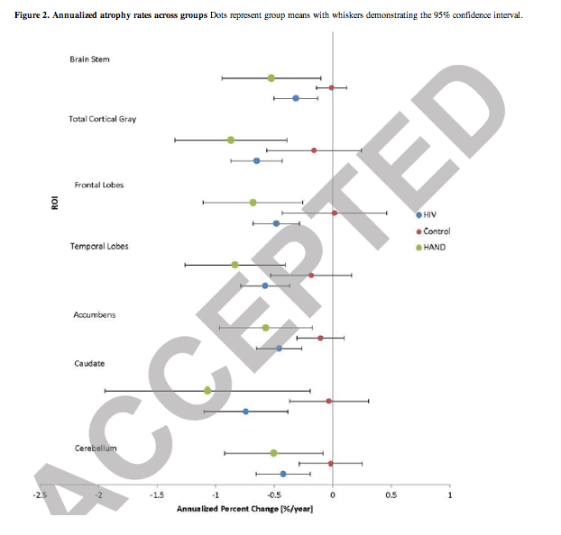
"The mean age of study participants was 63 years.....There is controversy as to whether plasma viral suppression is sufficient to halt detrimental brain changes with some arguing that volumetric reductions described in HIV are due to past injury. This study was designed to test whether plasma viral suppression is sufficient to halt progressive atrophy in older HIV-infected participants.....All had documented plasma viral suppression for each visit during longitudinal follow-up. Additionally, among the HIV-infected participants, 57% self reported persistent viral suppression for between 5-10 years, while 39% self-reported suppression for >10 years, although 17% reported either small "blips," or a planned drug holiday prior to enrollment, where their plasma HIV RNA was elevated for a short time but then returned to undetectable.......In longitudinal ROI models adjusted for age and sex, we uncovered progressive atrophy in the HIV-infected group exceeding rates seen among healthy controls (Table 2, bottom panel) with faster annualized rates of progressive atrophy in the cerebellum (0.42% vs 0.02%, p=0.016), caudate (0.74% vs 0.03%, p=0.012), frontal lobe (0.48% vs 0.01%, p=0.034), total cortical gray matter (0.65% vs 0.16%, p=0.027), brainstem (0.31% vs 0.01%, p=0.026), and pallidum (0.73% vs 0.39%, p=0.046) (Figure 1)......We add to a growing number of studies identifying detrimental neuropathogenic pathways, typically associated with persistent inflammation.
By virtue of the older age of our study participants compared to other studies, they may be particularly vulnerable to faster changes in brain atrophy as age-associated brain atrophy rates do not appear to have linear slopes in healthy aging.42 Furthermore, our data examined participants in age over 60 years, surpassing age set points thought to represent the 'tipping point' from linear to faster atrophy in presumed healthy aging. For example, a study of 1100 healthy elders noted nonlinear atrophy trajectories dominate in most subcortical regions after age 60 years.3
Cerebellar atrophy, white matter degeneration, and diffuse granule cell loss has been observed in postmortem studies of HIV-infected patients with cerebellar degeneration, cerebellar dysfunction, and gait ataxia.45
Our study investigated longitudinal rates of brain atrophy among older, HIV-infected individuals on suppressive antiretroviral therapy to determine if persistent viral control is sufficient to halt structural brain changes defined by prior studies where some participants had suboptimal viral suppression. Our cross-sectional analyses confirm limited volumetric reductions in HIV-infected participants with cognitive impairment compared to healthy controls, including regions previously noted to be affected in the setting of HIV, such as the cerebellum, nucleus accumbens and brainstem.36-38 These findings were driven by HIV-infected participants with HAND and we did not identify differences by HIV serostats alone. Among the HIV infected group, the baseline volume of the nucleus accumbens and its association to executive functioning is notable. A recent publication among HIV-uninfected adults found that the volume of the nucleus accumbens modulated the transition from Mild Cognitive Impairment to dementia of the Alzheimer's type disease.39 One could speculate parallels in HAND and Alzheimer's disease among older study participants as it demonstrates similarly affected subcortical structures.
Our primary hypotheses related to brain atrophy noted broad and sizably different longitudinal atrophy rates in HIV despite plasma viral suppression compared to age- and sex-matched cognitively normal and healthy controls. Faster atrophy rates were noted in the cerebellum, caudate, frontal lobe, total cortical gray matter, brainstem, and pallidum. HAND subjects additionally had greater atrophy rates in the temporal lobe and thalamus compared to controls. As some systematic variability in measures is expected with these technical approaches, we examined individual cases and ran sensitivity analyses excluding one individual with greater than 3 standard deviation change from the mean, retaining most of the primary findings.
Larger longitudinal volumetric reductions were noted in regions previously associated with HIV in cross-sectional studies which typically included some participants without viral suppression. Thus, there is some support that our findings may be due to HIV and HIV-related factors.13-15,19,21,23 The longitudinal nature of this work adds to extant literature, clarifying that reduction in brain volumes over time in HIV are not simply archival damage from the pre-cART era. This does not exclude the possibility that pre-cART brain changes set the stage for later amplified brain changes. Yet, other factors could have influenced these faster rates of compared to healthy controls, including HIV-independent factors such as smoking, cognitive reserve, noted by lower total educational attainment in table 1, and alcohol use as well as HIV-associated factors, including chronic inflammation, medication effects, and small vessel ischemic disease (SVID). A history of alcohol abuse was reported in 7/38 (18%) HIV infected participants with 6/38 (16%) reporting past abuse in substances besides alcohol (e.g. marijuana, cocaine, methamphetamine). Within the HIV-infected group, two participants (5.3%) reported having a history of both alcohol and other substance abuse. In comparison, only 2/24 (8.3%) controls reported past substance abuse with none attributed to alcohol. Given that participants in each group were excluded for current substance use disorders, these differences are more likely to inform baseline volumes than longitudinal change.
Most of our participants were infected with HIV in the 1980s, before cART was accessible, thus may differ from younger cohorts in brain vulnerability or survivor tendencies. A previous study conducting similar cross-sectional analysis confers HIV-infection in conjunction with aging is related to cortical volume loss .48 Our findings of no cross-sectional differences by serostatus alone was unexpected, suggesting the uniqueness of this sample of long-term survivors, although, differences emerged when examining those with HAND compared to controls. Nevertheless, we note progressive atrophy despite minimal baseline cross-sectional findings and can conclude that these changes are not limited to those with past injury. A lack of difference between HAND and non-HAND individuals further supports this contention. Our findings complement past work in this cohort noting Diffusion Tensor Imaging (DTI) deficits in white matter fiber integrity in older HIV participants compared to age-matched controls.24 Thus, we have identified multi-modal evidence of detrimental outcomes despite cART."
Figure 2. Annualized atrophy rates across groups Dots represent group means with whiskers demonstrating the 95% confidence interval.
------------------
Progressive Brain Atrophy Despite Persistent Viral Suppression in HIV Over Age 60.
Clifford, Katherine M. BA; Samboju, Vishal MS; Cobigo, Yann PhD; Milanini, Benedetta PhD; Marx, Gabriel A BA; Hellmuth, Joanna M. MD, MHS; Rosen, Howard J. MD; Kramer, Joel H.; Allen, Isabel E. PhD; Valcour, Victor G. MD, PhD
JAIDS June 22 2017
Background: Current HIV treatments are successful at suppressing plasma HIV RNA to undetectable levels for most adherent patients. Yet, emerging evidence suggests viral suppression will inadequately control inflammation and mitigate risk for progressive brain injury. We sought to quantify differences in longitudinal brain atrophy rates among older virally suppressed HIV-infected participants compared to that of healthy aging.
Methods: We examined longitudinal structural brain MRI atrophy rates employing region of interest assessments and voxel-wise tensor-based morphometry in HIV-infected participants over age 60 years (n=38) compared to age-matched HIV-uninfected healthy and cognitively normal controls (n=24).
Results: The mean age of participants was 63 years, the mean estimated duration of infection was 21 years and the median of duration of documented viral suppression was 3.2 years. Average proximal and nadir CD4 counts were 550 and 166, respectively; 15/38 (39%) met criteria for HIV-associated neurocognitive disorder. In models adjusting for age and sex, HIV serostatus was associated with more rapid average annualized rates of atrophy in the cerebellum (0.42% vs. 0.02%, p=0.016), caudate (0.74% vs. 0.03%, p=0.012), frontal lobe (0.48% vs. 0.01%, p=0.034), total cortical gray matter (0.65% vs. 0.16%, p=0.027), brain stem (0.31% vs. 0.01%, p=0.026), and pallidum (0.73% vs. 0.39%, p=0.046). Among those with HIV, atrophy rates did not differ statistically by cognitive status.
Conclusion: Despite persistent control of plasma viremia, these older HIV-infected participants demonstrate more rapid progressive brain atrophy when compared to healthy aging. Either HIV or other factors that differ between older HIV-infected participants and healthy controls could be responsible for these differences.
The literature is replete with cross-sectional analyses describing brain volumetric reductions associated with HIV serostatus, but do not typically examine rates of change and tend to include individuals not on cART or without plasma viral suppression. These reports often note _ENREF_19smaller subcortical and corpus callosum volumes and sometimes note associations to duration of HIV infection, plasma HIV RNA levels, or proximal and nadir CD4 T-lymphocyte counts.14-18 Collectively, these cross-sectional studies conclude HIV-associated effects that others note are independent of gray and white matter volumes from aging itself.14,19,20 These independent effects collectively decrease brain reserve, likely placing older HIV-infected patients at increased vulnerability to HAND and age-associated neurodegenerative disorders.
Several recent longitudinal studies examined volumetric changes among participants on cART with variable viral suppression, noting smaller white matter and gray matter volumes and expanded ventricular regions compared to HIV-uninfected controls. One study's sample varied in age from 21-55 years and only 54% were virally suppressed in plasma.16 In analyses focused on those with plasma suppression, widespread white matter atrophy was noted. Another study examined adults aged 20-67 years old where 20% were not on cART and mean plasma HIV RNA at baseline was 9,609 copies/ml, documenting increased rate of change in the lateral ventricles, insula, and hippocampus volume of HIV infected participants versus controls. Evidence of faster atrophy in the frontal, sensorimotor, and temporal-parietal cortex was also found in the HIV infected group.21 Another analysis conducted with Freesurfer over 26.6 months showed no longitudinal volume change in 21 virally suppressed (mean 53.9 years, range: 44-61). 22
The present work aimed to examine longitudinal rates of volumetric brain changes among HIV-infected older adults on cART maintaining persistent suppression of plasma HIV RNA compared to cognitively normal age- and sex-matched HIV-uninfected controls. There is controversy as to whether plasma viral suppression is sufficient to halt detrimental brain changes with some arguing that volumetric reductions described in HIV are due to past injury. This study was designed to test whether plasma viral suppression is sufficient to halt progressive atrophy in older HIV-infected participants. We employ region of interest (ROI) comparisons from neuroanatomy informed by prior studies. Secondarily, we employ Tensor Based Morphometry (TBM) at a voxel-wise level to explore volumetric reductions across all brain regions.
For these analyses, we selected only HIV-infected participants with documented persistently suppressed plasma HIV RNA defined as <400 copies/ml to accommodate for historical changes in lower level of detection during the enrollment period (05/2008 to 01/2016) and occasional viral blips of
unclear significance. Healthy controls were matched to the HIV-infected group by age and sex by including all healthy control males enrolled between the ages of 60 and 75 years and two randomly selected women to match the two HIV-infected women.
RESULTS - Among HIV-infected participants, 15/38 (39%) met research criteria for HAND, with four (11%) having Asymptomatic Neurocognitive Impairment and eleven (29%) having Mild Neurocognitive Disorder .35 All other HIV-infected and all healthy participants were cognitively normal by consensus conference.
On average HIV-infected reported 21 years since diagnosis range: 7-21.
Better executive functioning performance was associated with larger total baseline volume of deep gray structures, particularly, the caudate (p=0.005) and nucleus accumbens (p=0.001). Similarly, we found associations between executive function performance and frontal (p=0.017), temporal (p=0.006) and total cortical gray matter volumes (p =0.010) and a positive association between motor performance and both cerebellum volume (p=0.001) and thalamus (p=0.003).
Over the duration of follow-up , cognitive status remained stable for 66% of participants (n=25), whereas 5/15 participants with HAND no longer met HAND criteria at follow-up and 8/23 without HAND at baseline met criteria at follow-up, consistent with the fluctuating pattern described by others.35 As a group, neuropsychological performance, measured as a summary z-score of all tests administered, did not change over time (p=0.585).
In longitudinal ROI models adjusted for age and sex, we uncovered progressive atrophy in the HIV-infected group exceeding rates seen among healthy controls (Table 2, bottom panel) with faster annualized rates of progressive atrophy in the cerebellum (0.42% vs 0.02%, p=0.016), caudate (0.74% vs 0.03%, p=0.012), frontal lobe (0.48% vs 0.01%, p=0.034), total cortical gray matter (0.65% vs 0.16%, p=0.027), brainstem (0.31% vs 0.01%, p=0.026), and pallidum (0.73% vs 0.39%, p=0.046) (Figure 1).
We did not identify differences in longitudinal volume rates of change between those with compared to without HAND; however, the HAND group differed from controls with faster rates of progressive atrophy in the cerebellum (0.50% vs 0.02%, p=0.006), caudate (1.07% vs 0.03%, p=0.015), temporal lobes (0.83% vs 0.18%, p=0.049), total cortical gray matter (0.87% vs 0.16%, p=0.039), brainstem (0.52% vs 0.01%, p=0.005), pallidum (0.81% vs 0.39%, p=0.047), and thalamus (0.82% vs 0.44%, p=0.018) but not in total cerebral white matter (0.50% vs 0.55%, p=0.821). Analyses comparing controls to those without HAND did not identify any significant differences in atrophy rates.
|
|
| |
| |
|
|
|